The Visual System
1) Light
-each sensory organ is maximally sensitive to a certain physical stimulation -> sight (eye) to electromagnetic
radiation => particular form of radiation producing a visual response = light
-Newton: light = stream of particles, which are travelling in a straight line (early theory)
-each particle of light = photon -> intensity of light can be measured by counting the number of photons
=> NOTE: smallest intensity of light, which activates a receptor cell in the eye = 1 photon
-unlike Newton, Maxwell claimed that light acts as if it were a stream of WAVES
-> wavelength defines physical distance between peaks of photon waves (can vary between multiple kilometres to a
trillion of a centimetre) => HOWEVER: very short/long wavelengths are not visible
1) short wavelengths include gamma rays, X rays & ultraviolet rays
2) long wavelengths include electricity & broadcasting wavelengths associated with TV & radio
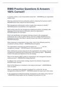
visible spectrum ranges from 380-760 nanometres (billionth of a meter) -> normal eyes see…
1) 400 nm as violet; 2) 500nm as blue-green; 3) 600nm as yellow-orange; 4) 700nm as red
-----------------------------------------------------------------------------------------------------------------------------------------------------------
2) Structure of Eye
-due to limited field of vision, humans can turn their heads & eyes
with great speed & accuracy => enabled by cooperative interaction
of extraocular muscles (6 per eye)
-eye is not made of rigid materials => maintains its shape due to
fluid pressure from within protected by orbital fat & eyelids
within its location (the orbit = depression within the skull)
blinking = prevents eye from drying out & keeps eye front clean
-hypothesis for why we don’t recognize that we can’t see while blinking:
-> when brain initiates blinking, it also prevents vision for a bit
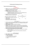
-3 different layers of eye (from outermost to innermost): 1) fibrous tunic; 2) vascular tunic; 3) retina
OUTERMOST, FIBROUS TUNIC
-sclera = membrane, which is outer covering (seen as the white) of the eye
-cornea = region, where the sclera bulges forward (1 st optically active element in the eye) -> simple, fixed lens that
gathers & concentrates light => due to forward-extension -> allows reception of light slightly behind obs.
=> anything that decreases its transparency reduces quality of images
MIDDLE, VASCULAR TUNIC
-mostly (2/3) consists of pigmented, spongy structure (choroid) -> main task: nourishes photoreceptors in retina
-toward front of eye, the middle layer curls away from wall of the eyeball -> forms slender structure (ciliary body)
=> manufactures aqueous humor = watery fluid filling anterior chamber (behind cornea)
provides lens & cornea with nourishment so that it doesn’t require own supply enables complete transparency
NOTE: aqueous fluid also helps to maintain shape of eye -> too little/much fluid => too much intraocular pressure
permanent increased intraocular pressure = glaucoma (most frequent cause of blinding eye disease)
-iris = colour membrane surrounding the central hole (actual colour varies from blue to black & is determined by
genetic makeup) -> controls amount of light entering
,-pupil = hole in the iris, through which the light enters -> its size is controlled by light reflex (Whytt’s reflex):
very small when light is bright (2mm) & very wide when light is dim (8mm)
-> adv of small pupil: 1) imperfections in the lens produce fewer distortions; 2) depth of focus (range of distances
over which objects are simultaneously in focus) is increased
=> NOTE: pupil size changes in response to emotional/attentional variables (greater pupil when highly interested)
-crystalline lens is located directly behind pupillary aperture + bc its curvature determines the amount of binding of
light, its shape is critical in bringing an image into focus at the rear of the eye (MAIN FUNCTION)
=> process of lens varying its focus = accommodation (changing focus by changing own shape):
normal shape = spherical (near objects in focus), BUT when ciliary muscles relax, the fluidal pressure flattens the
lens (brings far objects into focus)
*outer layer of lens never stops growing (always producing new protein fibres) they become more densely
packed in the centre of the lens with increasing age leading to sclerosis
*anything that reduces transparency of lens = cataract -> some are not impactful & others can lead to blindness
*lens is yellowy pigmented & thus not completely transparent -> density of yellow pigment increases with age
=> function of yellow pigment: screening out some of ultraviolet rays entering the eye + screening out some blue
(which often leads to discussion of people of different ages arguing for an object being green/blue)
-large chamber of the eye is filled with jelly-like substance (vitreous), which is generally clear = vitreous humor
INNERMOST, RETINA
-retina = screen of neural elements at the back of the eye (done by photoreceptors)
-> image formed by optical system is focused => ADDITIONALLY: light is changed/transduced into neural response
1) in daylight-active animals: retina is backed up light-absorbing dark layer (pigment epithelium)
-> PURPOSE: reducing amount of reflected/scattered light that could blur the final image
2) in nightlight-active animals: light that penetrates retina is reflected back through retina by shiny surface
(reflecting tapetum) => REASON: detection of light is more important than image clarity light passes through
retina twice, which doubles its intensity, BUT lowers image’s clarity
-3 major layers of neural tissue within the retina:
1) outermost layer (closest to scleral wall) -> contains photoreceptors => 2 types:
*long, thin, cylindrical cells = rods & *shorter, thicker, more tapered cells = cones
-> contain pigments that absorb light & start visual process (light reception)
=> 3 types of cells lay between photoreceptors & ganglion cells:
2) middle layer -> contains bipolar cells = neurons with 2 long-
extended processes
*1st making synapses with photoreceptors; *2 nd making
synapses with large ganglion cells (from 3rd layer)
=> function: transmission of modified signals from
photoreceptor cells to ganglion cells (the more glutamate is
produced in photoreceptors, the stronger the signal is sent
through bipolar cells)
NOTE: photoreceptors release glutamate inversely:
> low glutamate-release when receiving much light & vice versa
,-additionally, there’re 2 types of cells in the retina, which have lateral connections:
1) horizontal cells -> have short dendrites & connect with multiple adjacent photoreceptors
=> function: modifying the strength of the signals generated by neighbouring photoreceptors & giving feedback
about where signals may be stronger/weaker
2) amacrine cells -> interact with spatially adjacent ganglion cells
=> function: it is speculated that they receive & modify inputs from bipolar cells + have “switching function” in
controlling whether ganglion cells receive bipolar signals from cones or rods
-upside-down organization of retina (inverted orientation of cones & rods) forces light to pass retina first before
reaching photoreceptors => seems counterproductive, BUT is required because photoreceptors need oxygen supply,
which is guaranteed through being close to all the blood vessels
-in short:
1) neural signals are generated by photoreceptors
2) these neural signals are passed through a network of bipolar, amacrine & horizontal cells (that collect &
recombine the signals)
3) transformed signals are passed on to retinal ganglion cells (info on distribution of light is extracted & recoded)
4) this info is transmitted to the brain
FOVEA (SUB-PART OF RETINA)
-most important part of retina for perception is located around optic axis -> imaginary line from centre of retina to
centre of pupil -> beginning of optic axis is yellowish = macula lutea = yellow spot
-small, circular depression at the centre of the macula = fovea centralis -> critical in visual perception
=> when looking directly at an object, your eyes are rotated so that the image falls into the foveal region
-fovea has a very unique structure: photoreceptors are densely packed & there are only cones, which are longer &
thinner than in other regions => outside of fovea, the number of cones rapidly decreases & rods increase in number
RODS & CONES (DETAILED – CAN ALSO BE FOUND IN RETINA)
-main function of both: specifying light intensity falling on a particular photoreceptor
=> together with horizontal & amacrine cells all independent info are integrated and passed on to ganglion cells
-rods & cones have different functions -> based on finding that night-active animals have only rods & day-active
animals have only cones & humans (active during night and day) have both => proposed duplex retina theory
theory: there’re 2 separate visual systems
1) dependent on rods & responsible for vision under dim light conditions => scotopic vision
night blindness results from having no rods in retina
2) dependent on cones & responsible for vision under bright light conditions => photopic vision
day blindness results from having no cones in retina (looks completely different though: individual finds normal
levels of day-light painful, lacks colour vision & has poor visual acuity)
-additional findings:
*cones are highly concentrated at fovea (no rods found there) & are responsible for colour vision => 6 million
*rods more sensitive to low intensity-levels of light => 120 million
-rods & cones must interact with light first before they can signal its presence to the brain
=> they must absorb 1/more photons substance that absorbs light = called pigment
both rods & cones have visual pigments on the outside -> HOWEVER: pigmentation is different for both:
,1) rod pigments: rhodopsin = first isolated visual pigment -> consists of 2 parts:
*retinal = complex, organic molecule derived from vitamin A
*opsin = protein with large molecules that can potentially act as enzymes
=> when rhodopsin absorbs a photon of light, it changes shape & splits into its 2 parts
several enzymes are activated, which opens sodium channels
bc sodium ions flow into rod, rod cell undergoes hyperpolarization (negative charge becomes more negative)
to maintain sufficient rhodopsin supply, the process must be reversed: retinal & opsin regenerate rhodopsin
with the help of vitamin A & some enzymes
2) cone pigments: iodopsin = first isolated visual pigment -> breaks down to retinal (same as in rods) & photopsin
(different form of opsin in rods) when exposed to light => hyperpolarization (similar as before) stimulates bipolar
cells, which then in turn stimulate ganglion cells + complex interaction between both cell kinds occur, resulting in
axons of ganglion cells carrying neural signals towards the brain
GANGLION CELLS
-transmission of visual info from eye to brain: axons of retinal ganglion cells gather together & exit from eye through
a hole in retina & scleral wall => this resulting bundle of axons = optic nerve
-> blood vessels flow through centre of optic nerve & sustain metabolic needs of the eye
-because optic nerve must pass through retina, there are no photoreceptors in this region
-> no visual response possible at this spot (blind spot) => absence of vison
hole in visual field is automatically filled with information, fitting to its surrounding (visual completion)
-because neural axons form circular pattern, when leaving the eye -> this region is called optic disk
-nerve impulses transmitted to the brain via ganglion cell axons are already processed by the retina -> no raw data
=> integration of info from millions of adjacent photoreceptors by amacrine & horizontal cells
-----------------------------------------------------------------------------------------------------------------------------------------------------------
3) The Eye as an Optical Instrument
-to see, our eyes must capture light reflected from objects around us + pattern of light reaching the retina (retinal
image) must mirror the distribution of actual light distribution
=> THUS: quality of vision is dependent on how well light gets through to retina
-reflected light from the surface of objects is what our eyes pick up -> originally stems from emitted light (coming
from light bulb/sun) => objects’ surface usually absorbs large parts of the light, BUT reflect also parts of it
objects, which have high reflectance, appear light & objects, which have low reflectance, appear dark
-light’s way into our eyes: Cornea Anterior chamber Pupil and the lens the vitreous chamber Fovea
Retina (Inverted) [passes everything first & then reaches Photoreceptors horizontal cells bipolar cells
amacrine cells ganglion cells] optic nerve
NOTE: image is portrayed 180 degrees turned (left-right & top-bottom are reversed) later turned back by brain
-3 conditions of an object being seen must be fulfilled:
1) light must be sufficiently intense -> around 50% never reaches retina
2) distribution of light imaged on retina must be properly focused
3) spatial aspect must be preserved in the retinal image
,IMAGE FORMATION IN THE HUMAN EYE
-sharpness of images depends on 2 factors:
1) optical power of cornea & crystalline lens -> determined by accommodation ability
2) eyeball length from front to back
-Thomas Young: light behaves as if it consisted of waves -> light is divergent => HOWEVER: divergent light cannot be
focused something must reverse the divergence: convex lens
after hitting convex lens, 2 waves progressively come closer eventually they converge at focal point
for perfect vision: retina should be hit at focal point => emmetropic = eye having the perfect distance
SIGHT PROBLEMS
1) if eye is too short/if cornea bents light not sharply enough -> far-sighted = hypermetropia/hyperopia
=> focal point not yet reached at retina NOTE: accommodation process can solve mild distance-errors, BUT it
involves muscular effort & makes tired/leads to headaches quickly
alternative: placing a convex lens (glasses) in front of cornea, so that divergence is counterworked already
2) being near-sighted = myopia -> focal point reached somewhere before retina
=> NOTE: accommodation can’t help here 2 different types:
1) refractive myopia = abnormal shape of cornea -> light is bent too sharply
2) axial myopia = eyeball is too long -> too much time & space for the light to converge again
solution: concave lens, which leads to light diverging more
3) ability to refocus one’s lens relates to age -> very bad when we’re born & decreases after reaching age of 16y (due
to inner layers of lens dying resulting from refractory error (light-bending/focusing error) = presbyopia (old-sighted)
=> leads to increasing the near point distance (how close an object has to be brought to the eye until it can be no
longer held in focus) MEANING: older people need to hold books abnormally far to be able to focus on the letters
4) astigmatism = cornea is more sharply curved along 1 axis than it is along the other 1
-> result: cornea can’t sharply focus 2 different line orientations simultaneously on retina
=> can be corrected by lenses, which provide equal, opposite distortion
LASIK-surgery (re-sculpting cornea) is now an option for correcting everything but presbyopia
5) strabismus = eyes are not aligned with each other -> 1 is on-target, the other one is off-target
=> can lead to amblyopia = 1 eye is supressed & does not develop vision
6) Retinitis Pigmentosa (hereditary condition) = photoreceptors do not work like they are supposed to and a bundle
of tissue develops on your retina -> leads to blindness
7) age related macular degeneration = occurs most often in elderly people & constitutes of damage to the macula
-> results in a loss of vision in the centre of our visual field (NO total blindness) => genetic aspect involved
-----------------------------------------------------------------------------------------------------------------------------------------------------------
4) Neural Responses to Light (only read 4 & 5, but rely on 6)
RECEPTIVE FIELD OF A VISUAL NEURON
-info of a single ganglion cell represents the combined activity of a large number of rods & cones
-> THUS, a single ganglion cell responds to a light incident on a larger region of the retina (that cell’s receptive field)
=> MEANING: each ganglion cell processes info coming from a substantial zone of receptor cells in retina
, -sizes of receptive fields vary dependent on retinal location
-> very small close to the macula & becoming larger when moving further towards periphery of retina
=> with increasing eccentricity (deviation from the centre (fovea)) receptive fields increase in size
HOWEVER, the receptive field size can also vary for the same eccentricity (local variation)
-Hartline & Kuffler: when a single spot of light is displayed, a particular ganglion cell may react in 3 different ways:
1) on response -> burst of neural impulses right after onset of the stimulus
2) off response -> burst of neural impulses right after offset of the stimulus
3) on-off response -> burst of neural impulses right after onset & offset of the stimulus
=> particular ganglion cell response tends to vary depending on location of stimulus
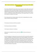
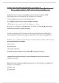
*organization of receptive field: some are on-centre receptive fields -> HOWEVER, off-centre receptive fields exist
too (approximately 50/50) => organization shown in graphic below
- the antagonistic regions of a receptive field (centre & surrounding)
compete with each other = lateral inhibition
=> enables to summarize the messages from many photoreceptors in
just 1 statement: “I have detected a light/dark boundary”
PARVO & MAGNO GANGLION CELLS
-classification system regarding ganglion cells’ size & shapes has been
established: most important difference = size -> parvo vs magno cells
*differences between parvo & magno cells:
1) parvo: as long as the same amount of illumination is present in centre & around, parvo cells do not distinguish
between different locations of illumination => neural response stays the same
responsible for stationary pattern analysis
2) magno: if location of same amount of illumination differs somehow (caused by object-movement), the neural
response varies responsible for motion detection
**other differences:
-sometimes, researchers add K cells to the categorization (others: P cells (80%) & M cells (10%))
-> relatively little research is done on them, BUT they more closely resemble P cells
=> HOWEVER, K cells have much better spatial resolution & react more sensitive to contrast compared to P cells
-----------------------------------------------------------------------------------------------------------------------------------------------------------
5) Perceptual Consequences of Centre/Surround Antagonism
1) Light
-each sensory organ is maximally sensitive to a certain physical stimulation -> sight (eye) to electromagnetic
radiation => particular form of radiation producing a visual response = light
-Newton: light = stream of particles, which are travelling in a straight line (early theory)
-each particle of light = photon -> intensity of light can be measured by counting the number of photons
=> NOTE: smallest intensity of light, which activates a receptor cell in the eye = 1 photon
-unlike Newton, Maxwell claimed that light acts as if it were a stream of WAVES
-> wavelength defines physical distance between peaks of photon waves (can vary between multiple kilometres to a
trillion of a centimetre) => HOWEVER: very short/long wavelengths are not visible
1) short wavelengths include gamma rays, X rays & ultraviolet rays
2) long wavelengths include electricity & broadcasting wavelengths associated with TV & radio
visible spectrum ranges from 380-760 nanometres (billionth of a meter) -> normal eyes see…
1) 400 nm as violet; 2) 500nm as blue-green; 3) 600nm as yellow-orange; 4) 700nm as red
-----------------------------------------------------------------------------------------------------------------------------------------------------------
2) Structure of Eye
-due to limited field of vision, humans can turn their heads & eyes
with great speed & accuracy => enabled by cooperative interaction
of extraocular muscles (6 per eye)
-eye is not made of rigid materials => maintains its shape due to
fluid pressure from within protected by orbital fat & eyelids
within its location (the orbit = depression within the skull)
blinking = prevents eye from drying out & keeps eye front clean
-hypothesis for why we don’t recognize that we can’t see while blinking:
-> when brain initiates blinking, it also prevents vision for a bit
-3 different layers of eye (from outermost to innermost): 1) fibrous tunic; 2) vascular tunic; 3) retina
OUTERMOST, FIBROUS TUNIC
-sclera = membrane, which is outer covering (seen as the white) of the eye
-cornea = region, where the sclera bulges forward (1 st optically active element in the eye) -> simple, fixed lens that
gathers & concentrates light => due to forward-extension -> allows reception of light slightly behind obs.
=> anything that decreases its transparency reduces quality of images
MIDDLE, VASCULAR TUNIC
-mostly (2/3) consists of pigmented, spongy structure (choroid) -> main task: nourishes photoreceptors in retina
-toward front of eye, the middle layer curls away from wall of the eyeball -> forms slender structure (ciliary body)
=> manufactures aqueous humor = watery fluid filling anterior chamber (behind cornea)
provides lens & cornea with nourishment so that it doesn’t require own supply enables complete transparency
NOTE: aqueous fluid also helps to maintain shape of eye -> too little/much fluid => too much intraocular pressure
permanent increased intraocular pressure = glaucoma (most frequent cause of blinding eye disease)
-iris = colour membrane surrounding the central hole (actual colour varies from blue to black & is determined by
genetic makeup) -> controls amount of light entering
,-pupil = hole in the iris, through which the light enters -> its size is controlled by light reflex (Whytt’s reflex):
very small when light is bright (2mm) & very wide when light is dim (8mm)
-> adv of small pupil: 1) imperfections in the lens produce fewer distortions; 2) depth of focus (range of distances
over which objects are simultaneously in focus) is increased
=> NOTE: pupil size changes in response to emotional/attentional variables (greater pupil when highly interested)
-crystalline lens is located directly behind pupillary aperture + bc its curvature determines the amount of binding of
light, its shape is critical in bringing an image into focus at the rear of the eye (MAIN FUNCTION)
=> process of lens varying its focus = accommodation (changing focus by changing own shape):
normal shape = spherical (near objects in focus), BUT when ciliary muscles relax, the fluidal pressure flattens the
lens (brings far objects into focus)
*outer layer of lens never stops growing (always producing new protein fibres) they become more densely
packed in the centre of the lens with increasing age leading to sclerosis
*anything that reduces transparency of lens = cataract -> some are not impactful & others can lead to blindness
*lens is yellowy pigmented & thus not completely transparent -> density of yellow pigment increases with age
=> function of yellow pigment: screening out some of ultraviolet rays entering the eye + screening out some blue
(which often leads to discussion of people of different ages arguing for an object being green/blue)
-large chamber of the eye is filled with jelly-like substance (vitreous), which is generally clear = vitreous humor
INNERMOST, RETINA
-retina = screen of neural elements at the back of the eye (done by photoreceptors)
-> image formed by optical system is focused => ADDITIONALLY: light is changed/transduced into neural response
1) in daylight-active animals: retina is backed up light-absorbing dark layer (pigment epithelium)
-> PURPOSE: reducing amount of reflected/scattered light that could blur the final image
2) in nightlight-active animals: light that penetrates retina is reflected back through retina by shiny surface
(reflecting tapetum) => REASON: detection of light is more important than image clarity light passes through
retina twice, which doubles its intensity, BUT lowers image’s clarity
-3 major layers of neural tissue within the retina:
1) outermost layer (closest to scleral wall) -> contains photoreceptors => 2 types:
*long, thin, cylindrical cells = rods & *shorter, thicker, more tapered cells = cones
-> contain pigments that absorb light & start visual process (light reception)
=> 3 types of cells lay between photoreceptors & ganglion cells:
2) middle layer -> contains bipolar cells = neurons with 2 long-
extended processes
*1st making synapses with photoreceptors; *2 nd making
synapses with large ganglion cells (from 3rd layer)
=> function: transmission of modified signals from
photoreceptor cells to ganglion cells (the more glutamate is
produced in photoreceptors, the stronger the signal is sent
through bipolar cells)
NOTE: photoreceptors release glutamate inversely:
> low glutamate-release when receiving much light & vice versa
,-additionally, there’re 2 types of cells in the retina, which have lateral connections:
1) horizontal cells -> have short dendrites & connect with multiple adjacent photoreceptors
=> function: modifying the strength of the signals generated by neighbouring photoreceptors & giving feedback
about where signals may be stronger/weaker
2) amacrine cells -> interact with spatially adjacent ganglion cells
=> function: it is speculated that they receive & modify inputs from bipolar cells + have “switching function” in
controlling whether ganglion cells receive bipolar signals from cones or rods
-upside-down organization of retina (inverted orientation of cones & rods) forces light to pass retina first before
reaching photoreceptors => seems counterproductive, BUT is required because photoreceptors need oxygen supply,
which is guaranteed through being close to all the blood vessels
-in short:
1) neural signals are generated by photoreceptors
2) these neural signals are passed through a network of bipolar, amacrine & horizontal cells (that collect &
recombine the signals)
3) transformed signals are passed on to retinal ganglion cells (info on distribution of light is extracted & recoded)
4) this info is transmitted to the brain
FOVEA (SUB-PART OF RETINA)
-most important part of retina for perception is located around optic axis -> imaginary line from centre of retina to
centre of pupil -> beginning of optic axis is yellowish = macula lutea = yellow spot
-small, circular depression at the centre of the macula = fovea centralis -> critical in visual perception
=> when looking directly at an object, your eyes are rotated so that the image falls into the foveal region
-fovea has a very unique structure: photoreceptors are densely packed & there are only cones, which are longer &
thinner than in other regions => outside of fovea, the number of cones rapidly decreases & rods increase in number
RODS & CONES (DETAILED – CAN ALSO BE FOUND IN RETINA)
-main function of both: specifying light intensity falling on a particular photoreceptor
=> together with horizontal & amacrine cells all independent info are integrated and passed on to ganglion cells
-rods & cones have different functions -> based on finding that night-active animals have only rods & day-active
animals have only cones & humans (active during night and day) have both => proposed duplex retina theory
theory: there’re 2 separate visual systems
1) dependent on rods & responsible for vision under dim light conditions => scotopic vision
night blindness results from having no rods in retina
2) dependent on cones & responsible for vision under bright light conditions => photopic vision
day blindness results from having no cones in retina (looks completely different though: individual finds normal
levels of day-light painful, lacks colour vision & has poor visual acuity)
-additional findings:
*cones are highly concentrated at fovea (no rods found there) & are responsible for colour vision => 6 million
*rods more sensitive to low intensity-levels of light => 120 million
-rods & cones must interact with light first before they can signal its presence to the brain
=> they must absorb 1/more photons substance that absorbs light = called pigment
both rods & cones have visual pigments on the outside -> HOWEVER: pigmentation is different for both:
,1) rod pigments: rhodopsin = first isolated visual pigment -> consists of 2 parts:
*retinal = complex, organic molecule derived from vitamin A
*opsin = protein with large molecules that can potentially act as enzymes
=> when rhodopsin absorbs a photon of light, it changes shape & splits into its 2 parts
several enzymes are activated, which opens sodium channels
bc sodium ions flow into rod, rod cell undergoes hyperpolarization (negative charge becomes more negative)
to maintain sufficient rhodopsin supply, the process must be reversed: retinal & opsin regenerate rhodopsin
with the help of vitamin A & some enzymes
2) cone pigments: iodopsin = first isolated visual pigment -> breaks down to retinal (same as in rods) & photopsin
(different form of opsin in rods) when exposed to light => hyperpolarization (similar as before) stimulates bipolar
cells, which then in turn stimulate ganglion cells + complex interaction between both cell kinds occur, resulting in
axons of ganglion cells carrying neural signals towards the brain
GANGLION CELLS
-transmission of visual info from eye to brain: axons of retinal ganglion cells gather together & exit from eye through
a hole in retina & scleral wall => this resulting bundle of axons = optic nerve
-> blood vessels flow through centre of optic nerve & sustain metabolic needs of the eye
-because optic nerve must pass through retina, there are no photoreceptors in this region
-> no visual response possible at this spot (blind spot) => absence of vison
hole in visual field is automatically filled with information, fitting to its surrounding (visual completion)
-because neural axons form circular pattern, when leaving the eye -> this region is called optic disk
-nerve impulses transmitted to the brain via ganglion cell axons are already processed by the retina -> no raw data
=> integration of info from millions of adjacent photoreceptors by amacrine & horizontal cells
-----------------------------------------------------------------------------------------------------------------------------------------------------------
3) The Eye as an Optical Instrument
-to see, our eyes must capture light reflected from objects around us + pattern of light reaching the retina (retinal
image) must mirror the distribution of actual light distribution
=> THUS: quality of vision is dependent on how well light gets through to retina
-reflected light from the surface of objects is what our eyes pick up -> originally stems from emitted light (coming
from light bulb/sun) => objects’ surface usually absorbs large parts of the light, BUT reflect also parts of it
objects, which have high reflectance, appear light & objects, which have low reflectance, appear dark
-light’s way into our eyes: Cornea Anterior chamber Pupil and the lens the vitreous chamber Fovea
Retina (Inverted) [passes everything first & then reaches Photoreceptors horizontal cells bipolar cells
amacrine cells ganglion cells] optic nerve
NOTE: image is portrayed 180 degrees turned (left-right & top-bottom are reversed) later turned back by brain
-3 conditions of an object being seen must be fulfilled:
1) light must be sufficiently intense -> around 50% never reaches retina
2) distribution of light imaged on retina must be properly focused
3) spatial aspect must be preserved in the retinal image
,IMAGE FORMATION IN THE HUMAN EYE
-sharpness of images depends on 2 factors:
1) optical power of cornea & crystalline lens -> determined by accommodation ability
2) eyeball length from front to back
-Thomas Young: light behaves as if it consisted of waves -> light is divergent => HOWEVER: divergent light cannot be
focused something must reverse the divergence: convex lens
after hitting convex lens, 2 waves progressively come closer eventually they converge at focal point
for perfect vision: retina should be hit at focal point => emmetropic = eye having the perfect distance
SIGHT PROBLEMS
1) if eye is too short/if cornea bents light not sharply enough -> far-sighted = hypermetropia/hyperopia
=> focal point not yet reached at retina NOTE: accommodation process can solve mild distance-errors, BUT it
involves muscular effort & makes tired/leads to headaches quickly
alternative: placing a convex lens (glasses) in front of cornea, so that divergence is counterworked already
2) being near-sighted = myopia -> focal point reached somewhere before retina
=> NOTE: accommodation can’t help here 2 different types:
1) refractive myopia = abnormal shape of cornea -> light is bent too sharply
2) axial myopia = eyeball is too long -> too much time & space for the light to converge again
solution: concave lens, which leads to light diverging more
3) ability to refocus one’s lens relates to age -> very bad when we’re born & decreases after reaching age of 16y (due
to inner layers of lens dying resulting from refractory error (light-bending/focusing error) = presbyopia (old-sighted)
=> leads to increasing the near point distance (how close an object has to be brought to the eye until it can be no
longer held in focus) MEANING: older people need to hold books abnormally far to be able to focus on the letters
4) astigmatism = cornea is more sharply curved along 1 axis than it is along the other 1
-> result: cornea can’t sharply focus 2 different line orientations simultaneously on retina
=> can be corrected by lenses, which provide equal, opposite distortion
LASIK-surgery (re-sculpting cornea) is now an option for correcting everything but presbyopia
5) strabismus = eyes are not aligned with each other -> 1 is on-target, the other one is off-target
=> can lead to amblyopia = 1 eye is supressed & does not develop vision
6) Retinitis Pigmentosa (hereditary condition) = photoreceptors do not work like they are supposed to and a bundle
of tissue develops on your retina -> leads to blindness
7) age related macular degeneration = occurs most often in elderly people & constitutes of damage to the macula
-> results in a loss of vision in the centre of our visual field (NO total blindness) => genetic aspect involved
-----------------------------------------------------------------------------------------------------------------------------------------------------------
4) Neural Responses to Light (only read 4 & 5, but rely on 6)
RECEPTIVE FIELD OF A VISUAL NEURON
-info of a single ganglion cell represents the combined activity of a large number of rods & cones
-> THUS, a single ganglion cell responds to a light incident on a larger region of the retina (that cell’s receptive field)
=> MEANING: each ganglion cell processes info coming from a substantial zone of receptor cells in retina
, -sizes of receptive fields vary dependent on retinal location
-> very small close to the macula & becoming larger when moving further towards periphery of retina
=> with increasing eccentricity (deviation from the centre (fovea)) receptive fields increase in size
HOWEVER, the receptive field size can also vary for the same eccentricity (local variation)
-Hartline & Kuffler: when a single spot of light is displayed, a particular ganglion cell may react in 3 different ways:
1) on response -> burst of neural impulses right after onset of the stimulus
2) off response -> burst of neural impulses right after offset of the stimulus
3) on-off response -> burst of neural impulses right after onset & offset of the stimulus
=> particular ganglion cell response tends to vary depending on location of stimulus
*organization of receptive field: some are on-centre receptive fields -> HOWEVER, off-centre receptive fields exist
too (approximately 50/50) => organization shown in graphic below
- the antagonistic regions of a receptive field (centre & surrounding)
compete with each other = lateral inhibition
=> enables to summarize the messages from many photoreceptors in
just 1 statement: “I have detected a light/dark boundary”
PARVO & MAGNO GANGLION CELLS
-classification system regarding ganglion cells’ size & shapes has been
established: most important difference = size -> parvo vs magno cells
*differences between parvo & magno cells:
1) parvo: as long as the same amount of illumination is present in centre & around, parvo cells do not distinguish
between different locations of illumination => neural response stays the same
responsible for stationary pattern analysis
2) magno: if location of same amount of illumination differs somehow (caused by object-movement), the neural
response varies responsible for motion detection
**other differences:
-sometimes, researchers add K cells to the categorization (others: P cells (80%) & M cells (10%))
-> relatively little research is done on them, BUT they more closely resemble P cells
=> HOWEVER, K cells have much better spatial resolution & react more sensitive to contrast compared to P cells
-----------------------------------------------------------------------------------------------------------------------------------------------------------
5) Perceptual Consequences of Centre/Surround Antagonism