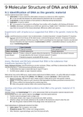
9 Molecular Structure of DNA and RNA
9.1 Identification of DNA as the genetic material
Genetic material must meet 4 criteria:
1. Information: it must contain the information necessary to construct an entire organism.
It must provide the blueprint for determining the inherited traits of an organism.
2. Transmission: it must be passed on from parents to offspring (during reproduction).
3. Replication: it must be copied.
To be passed on from parents to offspring+ from mother cell to daughter cells (during cell division).
4. Variation: phenotypic variability occurs within any species (e.g. height, color)-> therefore, it must also vary in
ways that can account for the known phenotypic differences within each species.
Experiments with streptococcus suggested that DNA is the genetic material (fig
9.1)
Griffith: used Pneumococcus strains+ mice to demonstrate a ‘transforming factor’; changes R bacteria S bacteria.
Fig 9.1d: extracts from tissues of the dead mouse: contained living type S bacteria-> living type R bacteria
alone couldn’t proliferate+ kill the mouse (fig. 9.1b) something from the dead type S bacteria transformed
the type R bacteria into type S bacteria= transformation (Griffith)-> unidentified substance causing this to
occur= transforming principle.
Type S (smooth) Type R (rough)
Capsule-secreting? Capsule-secreting Non-capsule-secreting
Colony morphology Smooth appearance Rough appearance
Virulence= ability to Infect a mouse capsule allows the bacteria to Destroyed by the animal’s immune
cause disease escape attack by the mouse’s immune system system
bacteria can grow+ eventually kill the mouse
Avery, Macleod, and McCarty showed that DNA is the substance that
transforms bacteria (fig 9.2)
Avery, Macleod+ McCarty: used Pneumococcus strains+ protease, DNase, and RNase to demonstrate DNA was the
transforming factor-> used biochemical purification procedures.
Purified DNA from type S mixed with type R: type R type S.
No DNA extract added: no type S bacterial colonies.
DNA extract may not be 100% pure; could contain small amount of RNA/ protein-> to verify: DNA extract samples
treated with enzymes that digest DNA (DNase), RNA (RNase), or protein (protease)-> DNA treated with:
RNase or protease: still type R type S; RNA/ protein in DNA extract wasn’t the genetic material.
DNase: no conversion type R type S bacteria; DNA degradation by DNase prevented conversion R S.
So they verified it by destroying the transforming factor.
DNA is the genetic material; DNA is the transforming principle.
Hershey and Chase provided evidence that DNA is the genetic material of T2
Phage
Hershey+ Chase: used bacteriophage T2+ E. coli to demonstrate DNA was the genetic material injected by the
bacteriophage into E. coli+ used to code for new phages= virus that injects bacteria.
Genetic material packaged inside a phage coat.
Molecularly simple; only 2 types of macromolecules: DNA+ proteins.
Infection: phage coat remains attached on the outside of the bacterium+ doesn’t enter the cell.
Only the genetic material (DNA) of the phage enters the bacterial cell.
Used radioisotopes to distinguish proteins from DNA:
Sulfur atoms (35S)= in proteins, not in DNA.
Phosphorus atoms (32P)= in DNA, not in phage proteins.
After infection: separated phage coats from the bacterial cells.
, 32P entered the bacterial cells; 35S remained outside the cells.
Genetic material of bacteriophages is DNA not proteins.
9.2 Overview of DNA and RNA structure
Friedrich Miescher: ‘discovery’ of DNA; identified a previously unknown phosphorus-containing substance that was
isolated from the nuclei of white blood cells; named this substance nuclein-> DNA+ RNA= nucleic acids.
Acidic molecules= releases hydrogen ions (H+) in solution+ have a net negative charge at neutral pH.
DNA+ RNA= macromolecules composed of smaller building blocks-> 4 levels of complexity (fig 9.3):
1. Nucleotides= form the repeating structural unit of nucleic acids.
2. Nucleotides are linked together in a linear manner to form a strand of DNA or RNA.
3. 2 strands of DNA (sometimes RNA) interact with each other to form a double helix.
4. 3D structure of DNA= from the folding+ bending of the double helix.
9.3 Nucleotide structure
Nucleotide= repeating structural unit of both DNA+ RNA-> has 3 components: min. 1 phosphate group, a pentose
sugar+ a nitrogenous base-> variety nucleotides; variety sugar+ nitrogenous bases (fig 9.4).
2 types of sugar: deoxyribose (DNA)+ ribose (RNA).
5 different bases (2 categories): purines+ pyrimidines.
Purine bases= contain a double-ring structure; adenine(A)+ guanine(G).
Pyrimidine bases= contain a single-ring structure; thymine(T), cytosine(C)+ uracil(U).
Thymine in DNA; uracil in RNA instead of thymine-> A, G+ U occur in both DNA+ RNA.
Bases+ sugars have a standard numbering system:
Nitrogen+ carbon atoms (in the ring structure of the bases):
numbers 1 – 9 (purines)+ 1 – 6 (pyrimidines).
5 carbons (in the sugars) numbers: have primes (1’ etc.)= to
distinguish them from the base numbers.
In sugar ring: carbons numbered in clockwise direction, beginning with a carbon next to the oxygen atom.
5th carbon is outside the ring structure.
In a single nucleotide: base always attached to the 1 ′ carbon atom+ 1/ more phosphate groups are attached
at the 5′ position-> -OH group attached to the 3′ carbon: important in allowing nucleotides to form covalent
linkages with each other.
Nucleoside= sugar+ base Nucleotide= phosphate(s)+ sugar+ base
Adenosine= ribose attached to adenine Adenosine monophosphate (AMP)= ribose,
Guanosine, cytidine+ uridine= ribose+ guanine, cytosine, or uracil adenine+ 1 phosphate
Deoxyadenosine, deoxyguanosine, deoxythymidine+ Adenosine triphosphate (ATP)= ribose, adenine+ 3
deoxycytidine= deoxyribose+ adenine, guanine, thymine, or phosphate groups
cytosine Phosphate attached to sugar via ester bond
9.4 Structure of a DNA strand
DNA (or RNA) nucleotides are linked together in a linear fashion (fig 9.7)-> few structural features:
Phosphodiester linkage= in DNA/ RNA strand; linkage that connects phosphate group to 2 sugar molecules.
Backbone= portion of DNA/ RNA strand that is composed of covalently linked phosphorates+ sugar.
Negatively charged due to a negative charge on each phosphate.
Bases project from the backbone.
Directionality= 5’ to 3’ orientation of nucleotides in a strand (DNA+ RNA).
In a strand: all sugar molecules are orientated in the same direction.
9.5 Discovery of the double helix
A few key events led to the discovery of the double-helix structure
MODEL BUILDING
Pauling: regions of proteins can fold into a secondary structure (α helix; fig 9.8a)