Musculoskeletal disorders
Introduction
In this assignment, the structure and function of the musculoskeletal system
will be understood to study the treatments available for disorders of the
system and their effectiveness and effective clinical management of common
musculoskeletal disorders.
(Figure 1 https://jpts.org.jo/wp-content/uploads/2017/03/musculoskeletal-
300x200.jpg)
Structure of the musculoskeletal system
Structure and identification of major bones, muscles, joints and supporting apparatus by visual examination of
diagrams or models and manipulative means in living subjects as appropriate.
Axial skeleton
The longitudinal axis of the body, which extends from the head to the bottom, forms an axial skeleton. It consists of:
The cranium (top part of the skull).
The maxilla (upper jaw bones).
The mandible (lower jaw bones).
The vertebral column (backbone).
Which comes down into various types of vertebrae like cervical, thorax, lumbar and, between them, the intervertebral
discs.
The rib cage and sternum (breastbone).
The cranium
The skull is made up of a series of plate-like bones joined by suture joints.
Together, they form the cranial cavity's bony base. The skull is made up of a
variety of bones bound by suture joints, resulting in a rigid space in which the
cerebrum and cerebellum are located.
(figure 2
https://upload.wikimedia.org/wikipedia/commons/thumb/8/8b/Axial_skeleton_diagram.svg/1200px-
Axial_skeleton_diagram.svg.png)
, (figure 3 https://resources.aofoundation.org/-/jssmedia/surgery/93/93_sb_d_i520.ashx?w=620)
The frontal bone - forming within the anterior part of the skull’s surface, as well as the superior part of the bony
section of the nose. It rests on top of the brain's frontal lobe. It houses the frontal air sinus, which can get clogged with
fluid if you have a sinus infection or congestion, and it also helps to lighten the frontal bone. The frontal bone also
serves as the floor of the anterior cranial fossa, which houses the brain's frontal lobe.
Sphenoid bone - The pituitary gland is housed within its shell, which is formed at the anterior and medial floor of the
middle cranial fossa (sella turcica). It has greater and lesser wings, with the greater wings forming the middle cranial
fossa's floor and the lesser wings forming the anterior cranial fossa's most posterior region. Many foramina in the bone
enable cranial nerves to exit the cranial cavity and innervate the eyes and face (optic via the optic canal, oculomotor
via the superior orbital fissure, trochlear via the superior orbital fissure, all three divisions of the trigeminal nerve, and
abducens via the superior orbital fissure). The greater wing of the sphenoid bone contributes a small portion of the
lateral surface of the skull. It connects to the frontal bone anteriorly, the temporal bone posteriorly, the parietal bone
via the lateral edge of its greater wings, and the clivus, the anterior superior projection of the occipital bone, which
houses the brainstem and basilar artery.
Temporal bones - Squamous and petrous portions of the bone are combined with (left and right sides). The exterior
surface of the skull is formed by the squamous portion, while the petrous portion houses the vestibulocochlear nerve
and its branches. The posterior two-thirds of the floor of the middle cranial fossa, as well as the anterior surfaces of
the posterior cranial fossa, are made up of these bones. The internal auditory meatus, a narrow opening that also
enables the facial nerve to exit the skull, allows the vestibulocochlear nerve to leave (move laterally away from
midline) the cranial vault. The vestibulocochlear nerve then splits into vestibular and cochlear branches in the inner
ear. The squamous part of the skull contributes a process to the zygomatic arch and forms the lateral inferior surface
of the skull. The temporal lobes of the brain are covered by the paired bones. At the posterior and anterior boundaries
of the bones, the jugular foramen and foramen lacerum are located.
, Occipital bone - The cerebellum resides in the cerebellar fossa of this bone, which arises at the most posterior surface
of the skull and overlies the occipital lobe. This bone comprises the foramen magnum. The inferior surface of the
occipital bone has occipital condyles that articulate with the Atlas (C1) vertebra. The largest, superior, medial, and
inferior nuchal lines are located on the occipital bone's most posterior surface. Layers of muscles that drive the neck
and back bind here.
The maxilla
The maxilla is the name for the upper jaw. The maxilla includes an air sinus, which helps
to lighten the bone and contributes to phonation tone. At the lateral margin of the bony
nasal septum, the maxilla articulates with the nasal bones, and at the lateral margin with
the zygomatic bone.
(Figure 4 https://thumbor.kenhub.com/-OyDtPg-7zxfX00wUG8FWzI1TcM=/fit-in/800x1600/filters:watermark(/
images/logo_url.png,-10,-10,0):background_color(FFFFFF):format(jpeg)/images/library/13967/Maxilla.png)
The mandible
The mandible is the name for the lower jaw. The temporal articular process articulates
with the mandibular condyle. The temporomandibular joint (TMJ) can protract and
withdraw the mandible, as well as raise and depress it. In the skull, the TMJ is the only
synovial joint. The mandible's ramus is the section below the condyle that forms the
posterior edge. The angle represents the mandible's corner, and the body represents the
jaw itself, where the teeth are attached. The inferior alveolar branch of the mandibular
nerve (a branch of the trigeminal nerve) enters the mandible and innervates the lower teeth through the mandibular
foramen, which is located on the internal surface of the ramus.
(figure 5 https://thumbor.kenhub.com/vJe2EfGS4ulCv4WzDXM-vj4tcgU=/fit-in/800x1600/filters:watermark(/
images/logo_url.png,-10,-10,0):background_color(FFFFFF):format(jpeg)/images/library/4972/
d2DFbcWqOqSTAXfJrVg3Mg_Foramen_mandibulae_01.png)
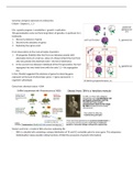
Vertebral column
Seven cervical vertebrae, twelve thoracic vertebrae, and five
lumbar vertebrae make up the spine. The sacrum, which is
usually made up of five fused vertebrae, and the coccyx, which
is usually made up of three or more fused vertebrae, are both
part of the vertebral column. Since each group of vertebrae has
its own anatomy, it has adapted to its purpose.
(figure 6)
(https://lh3.googleusercontent.com/proxy/C328gCTop26l5EO_S
0ifMKzPftP1QL0SozoUOG0PxPf9cVjjrMCvR4XgN6OXqTe7e
10qVbgsZpAz9W2F970WkDsz9Ul87ybIfcRnznnhvzGepUYwnRDWVJdmP51NJaNres47uwg7vJew-
4GHOgIfVKcqu9d3yAB6Eyf1m54kL8XWgx5kBjSABhu0aE)
Introduction
In this assignment, the structure and function of the musculoskeletal system
will be understood to study the treatments available for disorders of the
system and their effectiveness and effective clinical management of common
musculoskeletal disorders.
(Figure 1 https://jpts.org.jo/wp-content/uploads/2017/03/musculoskeletal-
300x200.jpg)
Structure of the musculoskeletal system
Structure and identification of major bones, muscles, joints and supporting apparatus by visual examination of
diagrams or models and manipulative means in living subjects as appropriate.
Axial skeleton
The longitudinal axis of the body, which extends from the head to the bottom, forms an axial skeleton. It consists of:
The cranium (top part of the skull).
The maxilla (upper jaw bones).
The mandible (lower jaw bones).
The vertebral column (backbone).
Which comes down into various types of vertebrae like cervical, thorax, lumbar and, between them, the intervertebral
discs.
The rib cage and sternum (breastbone).
The cranium
The skull is made up of a series of plate-like bones joined by suture joints.
Together, they form the cranial cavity's bony base. The skull is made up of a
variety of bones bound by suture joints, resulting in a rigid space in which the
cerebrum and cerebellum are located.
(figure 2
https://upload.wikimedia.org/wikipedia/commons/thumb/8/8b/Axial_skeleton_diagram.svg/1200px-
Axial_skeleton_diagram.svg.png)
, (figure 3 https://resources.aofoundation.org/-/jssmedia/surgery/93/93_sb_d_i520.ashx?w=620)
The frontal bone - forming within the anterior part of the skull’s surface, as well as the superior part of the bony
section of the nose. It rests on top of the brain's frontal lobe. It houses the frontal air sinus, which can get clogged with
fluid if you have a sinus infection or congestion, and it also helps to lighten the frontal bone. The frontal bone also
serves as the floor of the anterior cranial fossa, which houses the brain's frontal lobe.
Sphenoid bone - The pituitary gland is housed within its shell, which is formed at the anterior and medial floor of the
middle cranial fossa (sella turcica). It has greater and lesser wings, with the greater wings forming the middle cranial
fossa's floor and the lesser wings forming the anterior cranial fossa's most posterior region. Many foramina in the bone
enable cranial nerves to exit the cranial cavity and innervate the eyes and face (optic via the optic canal, oculomotor
via the superior orbital fissure, trochlear via the superior orbital fissure, all three divisions of the trigeminal nerve, and
abducens via the superior orbital fissure). The greater wing of the sphenoid bone contributes a small portion of the
lateral surface of the skull. It connects to the frontal bone anteriorly, the temporal bone posteriorly, the parietal bone
via the lateral edge of its greater wings, and the clivus, the anterior superior projection of the occipital bone, which
houses the brainstem and basilar artery.
Temporal bones - Squamous and petrous portions of the bone are combined with (left and right sides). The exterior
surface of the skull is formed by the squamous portion, while the petrous portion houses the vestibulocochlear nerve
and its branches. The posterior two-thirds of the floor of the middle cranial fossa, as well as the anterior surfaces of
the posterior cranial fossa, are made up of these bones. The internal auditory meatus, a narrow opening that also
enables the facial nerve to exit the skull, allows the vestibulocochlear nerve to leave (move laterally away from
midline) the cranial vault. The vestibulocochlear nerve then splits into vestibular and cochlear branches in the inner
ear. The squamous part of the skull contributes a process to the zygomatic arch and forms the lateral inferior surface
of the skull. The temporal lobes of the brain are covered by the paired bones. At the posterior and anterior boundaries
of the bones, the jugular foramen and foramen lacerum are located.
, Occipital bone - The cerebellum resides in the cerebellar fossa of this bone, which arises at the most posterior surface
of the skull and overlies the occipital lobe. This bone comprises the foramen magnum. The inferior surface of the
occipital bone has occipital condyles that articulate with the Atlas (C1) vertebra. The largest, superior, medial, and
inferior nuchal lines are located on the occipital bone's most posterior surface. Layers of muscles that drive the neck
and back bind here.
The maxilla
The maxilla is the name for the upper jaw. The maxilla includes an air sinus, which helps
to lighten the bone and contributes to phonation tone. At the lateral margin of the bony
nasal septum, the maxilla articulates with the nasal bones, and at the lateral margin with
the zygomatic bone.
(Figure 4 https://thumbor.kenhub.com/-OyDtPg-7zxfX00wUG8FWzI1TcM=/fit-in/800x1600/filters:watermark(/
images/logo_url.png,-10,-10,0):background_color(FFFFFF):format(jpeg)/images/library/13967/Maxilla.png)
The mandible
The mandible is the name for the lower jaw. The temporal articular process articulates
with the mandibular condyle. The temporomandibular joint (TMJ) can protract and
withdraw the mandible, as well as raise and depress it. In the skull, the TMJ is the only
synovial joint. The mandible's ramus is the section below the condyle that forms the
posterior edge. The angle represents the mandible's corner, and the body represents the
jaw itself, where the teeth are attached. The inferior alveolar branch of the mandibular
nerve (a branch of the trigeminal nerve) enters the mandible and innervates the lower teeth through the mandibular
foramen, which is located on the internal surface of the ramus.
(figure 5 https://thumbor.kenhub.com/vJe2EfGS4ulCv4WzDXM-vj4tcgU=/fit-in/800x1600/filters:watermark(/
images/logo_url.png,-10,-10,0):background_color(FFFFFF):format(jpeg)/images/library/4972/
d2DFbcWqOqSTAXfJrVg3Mg_Foramen_mandibulae_01.png)
Vertebral column
Seven cervical vertebrae, twelve thoracic vertebrae, and five
lumbar vertebrae make up the spine. The sacrum, which is
usually made up of five fused vertebrae, and the coccyx, which
is usually made up of three or more fused vertebrae, are both
part of the vertebral column. Since each group of vertebrae has
its own anatomy, it has adapted to its purpose.
(figure 6)
(https://lh3.googleusercontent.com/proxy/C328gCTop26l5EO_S
0ifMKzPftP1QL0SozoUOG0PxPf9cVjjrMCvR4XgN6OXqTe7e
10qVbgsZpAz9W2F970WkDsz9Ul87ybIfcRnznnhvzGepUYwnRDWVJdmP51NJaNres47uwg7vJew-
4GHOgIfVKcqu9d3yAB6Eyf1m54kL8XWgx5kBjSABhu0aE)