Human Pathology
3rd year biology
Theme 1: Neoplasia
Lecture 1.1 & 1.2: 25-1-2021
General principles of oncogenesis
Neoplasm does not always mean cancer
Neoplasia:
- “New growth” of cells
- Genetically driven!
- Benign, pre-malignant or malignant
A wart is also neoplasm of the skin
Different benign and malignant:
Benign neoplasm Malignant neoplasm
Only local growth Invasion in other tissues
No invasion in other tissue Can metastasize
No metastases Often rapid growth: mitoses, necrosis
(because blood vessels cannot keep up with
the rapid growth)
Often slowly growing Cellular atypia (look less like original cell,
big nuclei)
Cells without “atypia”
Neoplasia is genetically driven!
Types of genetic alterations:
- Mutations: a nucleotide change in DNA (e.g. substitution, insertion, deletion)
- Structural chromosomal alterations: translocations/gene fusions (part of chromosomes on other
chromosomes)
- Copy number variations: losses or gains of parts or whole chromosomes
- Viral transformation
3 types of genes can be involved in oncogenesis:
1. (proto)oncogenes
Function: promotion of cell proliferation and survival
Mutation/amplification: gain of function/ activation/ overexpression
2. Tumour suppressor genes
Function: inhibition of cell proliferation or induction of apoptosis
Mutation/loss: inactivation/ loss of function
3. Mutation/ mismatch repair genes
Function: repair of DNA replication errors
Mutation: loss of function (leads to an accumulation of DNA
errors)
Mutations can be somatic or germline
Somatic mutation:
- Acquired
- Present only in certain cells of the body, dependent on the cause
1
, Human Pathology
3rd year biology
- E.g. UV induced TP53 mutation in epithelial cells of the skin. Our caused by smoking etc.
Germline mutations:
- Hereditary, congenital
- Present in all cells of the body
Benign neoplasia’s usually have simple alterations
- Most common nevi (moles) have only one mutation
o 60% BRAF, 20% NRAS
- Spitz nevi (non-pigmented moles in children) have one chromosomal translocation
o ALK, ROS of NTRK translocation
- Some spitz nevi have one HRAS mutation and one gain of
chromosome 11p
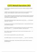
Cancer is a multistep process: clonal evolution model
- Several mutations (usually at least 6) in different genes
controlling cell growth, differentiation and death
- Often starting with a precancerous genetic change
- Additional genetic changes lead to cancerous growth
o Autonomous cell proliferation
o Loss of cell contact inhibition: invasion occurs
o New blood vessels develop
o Growth into vessels and metastasis can occur
Clonal evolution model: step by step. Every new mutation has a proliferation advantage/ survival
advantage.
General introduction to skin cancer
Skin is the most common primary location of cancer (52%)
Incidence of skin cancer is rising
Why is skin cancer that common?
- The skin is our largest organ
- Lifelong exposure to UV light, our skin is not made for Spanish sun
- The skin contains a lot of different structures that can give rise to different tumours
Different mutational processes generate unique combinations of mutation types, termed “Mutational
signatures”, e.g. when UV light is the cause, CC >>TT
2
, Human Pathology
3rd year biology
Basic principles of pathological examination
Routing patient with a skin tumour: First a skin biopsy is taken, then if necessary, a skin excision is done
(with a safety margin of normal skin).
Thin slices of the skin are made and diagnosed with a microscope by a dermatologist with a stain.
Histopathological investigations of skin tumours:
- Assessment of origin: from which tissue/cells is the tumour derived?
- Establish whether the tumour is benign of malignant (atypia, mitosis, necrosis, invasion)
Diagnosis
Pathological analysis of skin excision:
Diagnosis of a tumour:
- Routine histology is sufficient in most cases (HE staining) nuclei purple, cytoplasm pink
- Additional techniques are possible if needed
o Immunohistochemistry (10-15%)
o Molecular pathology (<5%)
Example primary skin tumour: in epidermis/keratinocyte
- Benign: wart
- Malignant: squamous cell carcinoma form keratin in cells
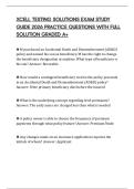
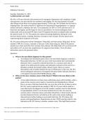
Immunohistochemistry: Usage of an antibody directed against an antigen on a (tumour) cell.
The binding is subsequently visualized with a chromogen
e.g. immunostaining on a slide: staining melanocytes by antibody
directed against MelanA
Molecular diagnostics: to establish/confirm diagnosis – for establish sensitivity for targeted therapy
- Analysis of DNA alterations in the tumour
o Mutations (by next generation sequencing)
o Translocations (by RNA fusion assay)
3
, Human Pathology
3rd year biology
o Copy number variations (by SNP array)
Example molecular diagnostics:
When you found out that the melanoma is caused by a BRAF mutation, the melanoma can then be treated
with BRAF inhibiting targeted therapy.
Skin tumours according to their origin (emphasis on their etiology/ oncogenesis)
Tumours are classified according to their origin
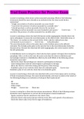
Epidermal tumours:
- Benign: wart
o Warts are caused by human papillomavirus (HPV)
o Hyperkeratinisation/ horn formation
HPV = DNA virus
o Infects squamous epithelium
o 170 types of which 40 genital transmitted
o HPV causes infected skin cells to multiply and form warts
Neoplasms caused by HPV:
o Warts (HPV 1,2,4,57)
o Genital warts (HPV 6 and 11)
o Squamous cell carcinoma of the skin (various HPV types,
in immunocompromised patients) in flat cells of
epidermis
o Genital SCC (HPV 6, 18, 31, 45)
o Oropharyngeal SCC (HPV 16)
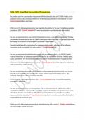
Malignant neoplasm derived from epidermis: Squamous cell carcinoma (SCC): clinical features
- Especially in areas with cumulative sun-exposition
- Can give rise to metastases: mostly nodal (2%) (lung and oesophagus also have squamous cells)
Histology: you can see desmosomes between cells (with large nuclei) and keratinization
4
3rd year biology
Theme 1: Neoplasia
Lecture 1.1 & 1.2: 25-1-2021
General principles of oncogenesis
Neoplasm does not always mean cancer
Neoplasia:
- “New growth” of cells
- Genetically driven!
- Benign, pre-malignant or malignant
A wart is also neoplasm of the skin
Different benign and malignant:
Benign neoplasm Malignant neoplasm
Only local growth Invasion in other tissues
No invasion in other tissue Can metastasize
No metastases Often rapid growth: mitoses, necrosis
(because blood vessels cannot keep up with
the rapid growth)
Often slowly growing Cellular atypia (look less like original cell,
big nuclei)
Cells without “atypia”
Neoplasia is genetically driven!
Types of genetic alterations:
- Mutations: a nucleotide change in DNA (e.g. substitution, insertion, deletion)
- Structural chromosomal alterations: translocations/gene fusions (part of chromosomes on other
chromosomes)
- Copy number variations: losses or gains of parts or whole chromosomes
- Viral transformation
3 types of genes can be involved in oncogenesis:
1. (proto)oncogenes
Function: promotion of cell proliferation and survival
Mutation/amplification: gain of function/ activation/ overexpression
2. Tumour suppressor genes
Function: inhibition of cell proliferation or induction of apoptosis
Mutation/loss: inactivation/ loss of function
3. Mutation/ mismatch repair genes
Function: repair of DNA replication errors
Mutation: loss of function (leads to an accumulation of DNA
errors)
Mutations can be somatic or germline
Somatic mutation:
- Acquired
- Present only in certain cells of the body, dependent on the cause
1
, Human Pathology
3rd year biology
- E.g. UV induced TP53 mutation in epithelial cells of the skin. Our caused by smoking etc.
Germline mutations:
- Hereditary, congenital
- Present in all cells of the body
Benign neoplasia’s usually have simple alterations
- Most common nevi (moles) have only one mutation
o 60% BRAF, 20% NRAS
- Spitz nevi (non-pigmented moles in children) have one chromosomal translocation
o ALK, ROS of NTRK translocation
- Some spitz nevi have one HRAS mutation and one gain of
chromosome 11p
Cancer is a multistep process: clonal evolution model
- Several mutations (usually at least 6) in different genes
controlling cell growth, differentiation and death
- Often starting with a precancerous genetic change
- Additional genetic changes lead to cancerous growth
o Autonomous cell proliferation
o Loss of cell contact inhibition: invasion occurs
o New blood vessels develop
o Growth into vessels and metastasis can occur
Clonal evolution model: step by step. Every new mutation has a proliferation advantage/ survival
advantage.
General introduction to skin cancer
Skin is the most common primary location of cancer (52%)
Incidence of skin cancer is rising
Why is skin cancer that common?
- The skin is our largest organ
- Lifelong exposure to UV light, our skin is not made for Spanish sun
- The skin contains a lot of different structures that can give rise to different tumours
Different mutational processes generate unique combinations of mutation types, termed “Mutational
signatures”, e.g. when UV light is the cause, CC >>TT
2
, Human Pathology
3rd year biology
Basic principles of pathological examination
Routing patient with a skin tumour: First a skin biopsy is taken, then if necessary, a skin excision is done
(with a safety margin of normal skin).
Thin slices of the skin are made and diagnosed with a microscope by a dermatologist with a stain.
Histopathological investigations of skin tumours:
- Assessment of origin: from which tissue/cells is the tumour derived?
- Establish whether the tumour is benign of malignant (atypia, mitosis, necrosis, invasion)
Diagnosis
Pathological analysis of skin excision:
Diagnosis of a tumour:
- Routine histology is sufficient in most cases (HE staining) nuclei purple, cytoplasm pink
- Additional techniques are possible if needed
o Immunohistochemistry (10-15%)
o Molecular pathology (<5%)
Example primary skin tumour: in epidermis/keratinocyte
- Benign: wart
- Malignant: squamous cell carcinoma form keratin in cells
Immunohistochemistry: Usage of an antibody directed against an antigen on a (tumour) cell.
The binding is subsequently visualized with a chromogen
e.g. immunostaining on a slide: staining melanocytes by antibody
directed against MelanA
Molecular diagnostics: to establish/confirm diagnosis – for establish sensitivity for targeted therapy
- Analysis of DNA alterations in the tumour
o Mutations (by next generation sequencing)
o Translocations (by RNA fusion assay)
3
, Human Pathology
3rd year biology
o Copy number variations (by SNP array)
Example molecular diagnostics:
When you found out that the melanoma is caused by a BRAF mutation, the melanoma can then be treated
with BRAF inhibiting targeted therapy.
Skin tumours according to their origin (emphasis on their etiology/ oncogenesis)
Tumours are classified according to their origin
Epidermal tumours:
- Benign: wart
o Warts are caused by human papillomavirus (HPV)
o Hyperkeratinisation/ horn formation
HPV = DNA virus
o Infects squamous epithelium
o 170 types of which 40 genital transmitted
o HPV causes infected skin cells to multiply and form warts
Neoplasms caused by HPV:
o Warts (HPV 1,2,4,57)
o Genital warts (HPV 6 and 11)
o Squamous cell carcinoma of the skin (various HPV types,
in immunocompromised patients) in flat cells of
epidermis
o Genital SCC (HPV 6, 18, 31, 45)
o Oropharyngeal SCC (HPV 16)
Malignant neoplasm derived from epidermis: Squamous cell carcinoma (SCC): clinical features
- Especially in areas with cumulative sun-exposition
- Can give rise to metastases: mostly nodal (2%) (lung and oesophagus also have squamous cells)
Histology: you can see desmosomes between cells (with large nuclei) and keratinization
4