

242 Med Surg
Exam 2
Unit 4-6
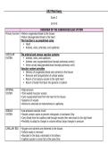
OVERVIEW OF THE CARDIOVASCULAR SYSTEM
Primary function •Deliver oxygenated blood to the tissues
•Return deoxygenated blood to the heart
This function is accomplished using
Heart
Arteries, veins, arterioles, and capillaries
VASCULAR The arterial and venous vascular systems
SYSTEM Arteries, veins, and capillaries
Arteries carry oxygenated blood (except pulmonary artery)
Veins convey deoxygenated blood (except pulmonary vein)
Vascular system provides
Delivery of oxygenated blood and nutrients to the tissues
Removal and transportation of cellular wastes
Return of circulatory volume to the right heart
Return of lymph fluid back into general circulation
ARTERIAL •High-pressure
SYSTEM •Thick-walled muscular vessels
•Carry oxygenated blood from the heart to the tissues
•Sequence of vessels
•Arteries to arterioles to metarterioles to capillaries
VENOUS •Low-pressure vascular circuit
SYSTEM •Vessels contain valves to prevent retrograde, or backward, flow
•Carry blood from the capillary bed through venules then veins back to the right heart
•Flexibility to adapt to changes in volume without large changes in pressure
CAPILLARY BED •Oxygen and nutrients are delivered to the tissues
•Cellular waste is removed
•Abundant in the body, estimated in the billions
•Capillary system is never full at the same time
,•When precapillary sphincters are open
•Capillaries are filled with blood
•When a precapillary sphincter closes
•Corresponding capillary shuts down
ANATOMY OF THE HEART
•Pericardium consists of 2 layers
•Outer layer
•Inner layer
•Heart muscle is composed of 3 layers
•Epicardium
•Myocardium
•Endocardium
•4 heart chambers
Right and left atria
Right and left ventricles
•Valves
Atrioventricular (AV) valves
Semilunar valves
•Blood supply
Left and right coronary arteries supply blood to the heart
A – ATRIAL
P – PULMONIC
E – ERBS POINT
TO – TRICUSPID
ME - MITRAL
,Murmur •Valve stenosis
•Valve regurgitation of insufficiency
•Valve prolapse
•Endocarditis
•Pericarditis
•Septal defects
•Atrial or ventricular
Coronary •The coronary arteries feed the myocardium of the heart.
Circulation •The blockages in the coronary arteries and their branches will lead to a MI.
CARDIAC Heart rate (HR) - Can be affected by many variables
OUTPUT Stroke Volume (SV) -
CO = Preload
Afterload
HR x SV Contractility
DIAGNOSTIC •Laboratory markers as predictors of heart disease
ASSESSMENT •Laboratory markers of acute cardiac damage or injury
•Diagnostic imaging studies
•Radiology
•Echocardiography
•Cardiac stress testing
Catheterization
•Right heart catheterization through a suitable vein (femoral, brachial,
subclavian)
•Left heart catheterization through a suitable artery (femoral, brachial,
radial)
•Atropine at bedside (in case of Bradycardia)
Angiography
•Primary reason cardiac catheterization is performed
, ELECTROCARDIOGRA Electrodes on extremities and 6 chest locations
M (ECG) Produce a picture of cardiac electrical activity
Small block = 0.04 sec
Large block = 0.2 sec (5 small blocks)
Principal waveforms
P wave, QRS complex, and T wave
Principal segments
PR segment and ST segment
Intervals
PR interval, QRS interval, and QT interval
Exam 2
Unit 4-6
OVERVIEW OF THE CARDIOVASCULAR SYSTEM
Primary function •Deliver oxygenated blood to the tissues
•Return deoxygenated blood to the heart
This function is accomplished using
Heart
Arteries, veins, arterioles, and capillaries
VASCULAR The arterial and venous vascular systems
SYSTEM Arteries, veins, and capillaries
Arteries carry oxygenated blood (except pulmonary artery)
Veins convey deoxygenated blood (except pulmonary vein)
Vascular system provides
Delivery of oxygenated blood and nutrients to the tissues
Removal and transportation of cellular wastes
Return of circulatory volume to the right heart
Return of lymph fluid back into general circulation
ARTERIAL •High-pressure
SYSTEM •Thick-walled muscular vessels
•Carry oxygenated blood from the heart to the tissues
•Sequence of vessels
•Arteries to arterioles to metarterioles to capillaries
VENOUS •Low-pressure vascular circuit
SYSTEM •Vessels contain valves to prevent retrograde, or backward, flow
•Carry blood from the capillary bed through venules then veins back to the right heart
•Flexibility to adapt to changes in volume without large changes in pressure
CAPILLARY BED •Oxygen and nutrients are delivered to the tissues
•Cellular waste is removed
•Abundant in the body, estimated in the billions
•Capillary system is never full at the same time
,•When precapillary sphincters are open
•Capillaries are filled with blood
•When a precapillary sphincter closes
•Corresponding capillary shuts down
ANATOMY OF THE HEART
•Pericardium consists of 2 layers
•Outer layer
•Inner layer
•Heart muscle is composed of 3 layers
•Epicardium
•Myocardium
•Endocardium
•4 heart chambers
Right and left atria
Right and left ventricles
•Valves
Atrioventricular (AV) valves
Semilunar valves
•Blood supply
Left and right coronary arteries supply blood to the heart
A – ATRIAL
P – PULMONIC
E – ERBS POINT
TO – TRICUSPID
ME - MITRAL
,Murmur •Valve stenosis
•Valve regurgitation of insufficiency
•Valve prolapse
•Endocarditis
•Pericarditis
•Septal defects
•Atrial or ventricular
Coronary •The coronary arteries feed the myocardium of the heart.
Circulation •The blockages in the coronary arteries and their branches will lead to a MI.
CARDIAC Heart rate (HR) - Can be affected by many variables
OUTPUT Stroke Volume (SV) -
CO = Preload
Afterload
HR x SV Contractility
DIAGNOSTIC •Laboratory markers as predictors of heart disease
ASSESSMENT •Laboratory markers of acute cardiac damage or injury
•Diagnostic imaging studies
•Radiology
•Echocardiography
•Cardiac stress testing
Catheterization
•Right heart catheterization through a suitable vein (femoral, brachial,
subclavian)
•Left heart catheterization through a suitable artery (femoral, brachial,
radial)
•Atropine at bedside (in case of Bradycardia)
Angiography
•Primary reason cardiac catheterization is performed
, ELECTROCARDIOGRA Electrodes on extremities and 6 chest locations
M (ECG) Produce a picture of cardiac electrical activity
Small block = 0.04 sec
Large block = 0.2 sec (5 small blocks)
Principal waveforms
P wave, QRS complex, and T wave
Principal segments
PR segment and ST segment
Intervals
PR interval, QRS interval, and QT interval

