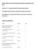
ANCC Cardiac-Vascular Nursing Certification Examination (CV-
BC™)
Version 2.0 — Advanced Clinical Practice Question Bank
150 Multiple-Choice Questions | Detailed & Complicated Scenarios
Hard Difficulty | For Experienced Cardiac-Vascular Nurses & Advanced
Practitioners
TABLE OF CONTENTS
Domain Questions Page
Domain I: Advanced Assessment &
36 2
Diagnostic Mastery (Q1–36)
└─ High-Fidelity Hemodynamics &
Q1–Q9 2
Waveform Analysis
└─ Advanced Cardiac Imaging & Biomarker
Q10–Q18 3
Interpretation
└─ Complex ECG & Electrophysiological
Q19–Q27 5
Phenomena
└─ Invasive & Non-Invasive Vascular
Q28–Q36 6
Diagnostics
Domain II: Complex Planning & High-Acuity
54 7
Implementation (Q37–90)
└─ High-Risk ACS & Shock States Q37–Q48 7
,Domain Questions Page
└─ Advanced Heart Failure, MCS &
Q49–Q62 9
Transplant
└─ Refractory Arrhythmias & Device Therapy Q63–Q74 11
└─ Complex Valvular, Structural & Aortic
Q75–Q82 13
Disease
└─ Pharmacogenomics, Polypharmacy &
Q83–Q90 14
Drug Toxicity
Domain III: Evaluation, Modification &
30 15
Adverse Event Forensics (Q91–120)
└─ Procedural Complications & Sentinel
Q91–Q102 15
Events
└─ Hemodynamic Deterioration & Rescue
Q103–Q112 17
Therapies
└─ Post-Operative Critical Care & Recovery
Q113–Q120 18
Trajectory
Domain IV: Patient Empowerment,
30 19
Community Health & Ethics (Q121–150)
└─ Shared Decision-Making & End-of-Life Q121–Q130 19
└─ Genomic, Lifestyle & Population Health Q131–Q140 21
└─ Advanced Professional Role &
Q141–Q150 22
Medicolegal Nuances
DOMAIN I: ADVANCED ASSESSMENT & DIAGNOSTIC MASTERY
,Subdomain: High-Fidelity Hemodynamics & Waveform Analysis
🟢 Q1. A patient with a pulmonary artery catheter displays a pulmonary artery pressure (PAP)
waveform that transitions from a normal systolic peak to an elevated diastolic plateau. The mean PAP
is 42 mm Hg, and the pulmonary capillary wedge pressure (PCWP) is 12 mm Hg. This transpulmonary
gradient (TPG) of 30 mm Hg is most consistent with which of the following pathophysiological states,
and what additional calculation is required to confirm the diagnosis?
A. Isolated left-sided heart failure; calculate pulmonary vascular resistance (PVR)
B. Combined pre- and post-capillary pulmonary hypertension; calculate diastolic pressure gradient
(DPG)
C. Reactive pulmonary vasoconstriction from ARDS; calculate the pulmonary artery pulsatility index
(PAPi)
D. Chronic thromboembolic disease; calculate the pulmonary acceleration time
🔴🔴 Correct Answer: B
Rationale: A TPG > 12 mm Hg with a PCWP ≤ 15 mm Hg suggests pre-capillary PH. In the presence of
elevated PCWP (> 15), an elevated TPG defines combined pre- and post-capillary PH (Cpc-PH). The
diastolic pressure gradient (DPG = PA diastolic – PCWP) > 7 mm Hg confirms Cpc-PH, which is distinct
from isolated post-capillary PH (DPG < 7). Option A ignores the elevated TPG. Option C is not specific.
Option D is a non-invasive echo parameter.
🟢 Q2. During an intra-aortic balloon pump (IABP) weaning trial, the patient's arterial waveform shows
a 1:2 augmentation ratio with a diastolic augmentation pressure that is 90% of the systolic pressure.
The augmented diastolic pressure is measured at 105 mm Hg while the unassisted systolic pressure is
110 mm Hg. This set of numbers indicates:
A. Optimal IABP timing with excellent afterload reduction
B. Suboptimal timing with early inflation causing increased afterload
C. 1:2 weaning mode with adequate hemodynamic support but borderline augmentation
D. Late inflation causing decreased coronary perfusion
🔴🔴 Correct Answer: C
Rationale: IABP weaning often uses a 1:2 mode (every other beat). A diastolic augmentation pressure
that is 90% of the unassisted systolic pressure (here 105 vs 110) is generally acceptable (typically target
> 80%). This indicates adequate support, but the patient is being weaned. Option A would be 1:1
mode. Option B would show elevated systolic pressure with inflation. Option D would show a dip or
loss of augmentation.
🟢 Q3. A patient with septic cardiomyopathy has a cardiac output measured by thermodilution
(COTD) of 4.2 L/min and a simultaneous continuous cardiac output (CCO) via a pulmonary artery
catheter showing 6.8 L/min. A right atrial injection of cold saline confirms thermodilution accuracy.
Which of the following best explains the discrepancy?
A. The presence of severe tricuspid regurgitation causing thermodilution underestimation
B. A rapid heart rate causing loss of indicator
C. An intrapulmonary shunt causing indicator loss
, D. A low stroke volume variation causing overestimation of continuous CO
🔴🔴 Correct Answer: A
Rationale: Severe tricuspid regurgitation causes the cold injectate to recirculate across the tricuspid
valve, leading to a prolonged thermodilution curve and underestimation of cardiac output by bolus
thermodilution. CCO (via a heated filament) may be less affected. Option B is incorrect (rapid rates can
cause underestimation or overestimation depending on the algorithm, but TR is a classic cause). C and
D are less likely.
🟢 Q4. A patient with a continuous-flow LVAD (HeartMate 3) has an arterial waveform that shows a
mean arterial pressure (MAP) of 85 mm Hg by Doppler, but an invasive radial artery line shows a MAP
of 72 mm Hg. The LVAD speed is 5600 RPM and power is 5.2 W. What is the most likely cause of the
discrepancy and the appropriate nursing action?
A. Overestimation by Doppler due to arterial calcification; adjust the LVAD speed down
B. Underestimation by radial line due to damped waveform from low pulsatility; use the Doppler MAP
for management
C. Inflow cannula obstruction causing power spikes; prepare for pump exchange
D. Aortic valve opening causing systemic pressure overshoot; increase afterload reduction
🔴🔴 Correct Answer: B
Rationale: In continuous-flow LVAD patients, the systemic arterial waveform is often under-damped or
damped due to low pulsatility, making radial arterial lines inaccurate. Doppler MAP (using the formula:
[SBP + 2×DBP]/3) is the preferred and more accurate method for titrating vasoactive medications.
Option A is wrong because Doppler is accurate; calcification affects ABI, not MAP. C and D are not
supported by the data.
🟢 Q5. A central venous pressure (CVP) waveform shows a dominant "V" wave and a rapid "Y"
descent, with a mean CVP of 18 mm Hg. The patient is in sinus rhythm with a normal PR interval. This
waveform is most specific for:
A. Tricuspid stenosis with elevated right atrial pressure
B. Severe tricuspid regurgitation with right ventricular volume overload
C. Cardiac tamponade with pulsus paradoxus
D. Restrictive cardiomyopathy with constrictive physiology
🔴🔴 Correct Answer: B
Rationale: A dominant V wave in the CVP with a steep Y descent is the classic waveform for tricuspid
regurgitation. The V wave represents systolic filling of the right atrium due to regurgitant flow, and the
Y descent is rapid passive emptying into the right ventricle. Option A (tricuspid stenosis) shows a
prominent A wave and blunted Y descent. C and D show blunted or different patterns.
🟢 Q6. A patient with cardiogenic shock has a pulmonary artery pulsatility index (PAPi) of 0.8. The
formula for PAPi is (PASP – PADP) / mean RAP. What does this value indicate regarding right
ventricular function and what intervention is most appropriate?
BC™)
Version 2.0 — Advanced Clinical Practice Question Bank
150 Multiple-Choice Questions | Detailed & Complicated Scenarios
Hard Difficulty | For Experienced Cardiac-Vascular Nurses & Advanced
Practitioners
TABLE OF CONTENTS
Domain Questions Page
Domain I: Advanced Assessment &
36 2
Diagnostic Mastery (Q1–36)
└─ High-Fidelity Hemodynamics &
Q1–Q9 2
Waveform Analysis
└─ Advanced Cardiac Imaging & Biomarker
Q10–Q18 3
Interpretation
└─ Complex ECG & Electrophysiological
Q19–Q27 5
Phenomena
└─ Invasive & Non-Invasive Vascular
Q28–Q36 6
Diagnostics
Domain II: Complex Planning & High-Acuity
54 7
Implementation (Q37–90)
└─ High-Risk ACS & Shock States Q37–Q48 7
,Domain Questions Page
└─ Advanced Heart Failure, MCS &
Q49–Q62 9
Transplant
└─ Refractory Arrhythmias & Device Therapy Q63–Q74 11
└─ Complex Valvular, Structural & Aortic
Q75–Q82 13
Disease
└─ Pharmacogenomics, Polypharmacy &
Q83–Q90 14
Drug Toxicity
Domain III: Evaluation, Modification &
30 15
Adverse Event Forensics (Q91–120)
└─ Procedural Complications & Sentinel
Q91–Q102 15
Events
└─ Hemodynamic Deterioration & Rescue
Q103–Q112 17
Therapies
└─ Post-Operative Critical Care & Recovery
Q113–Q120 18
Trajectory
Domain IV: Patient Empowerment,
30 19
Community Health & Ethics (Q121–150)
└─ Shared Decision-Making & End-of-Life Q121–Q130 19
└─ Genomic, Lifestyle & Population Health Q131–Q140 21
└─ Advanced Professional Role &
Q141–Q150 22
Medicolegal Nuances
DOMAIN I: ADVANCED ASSESSMENT & DIAGNOSTIC MASTERY
,Subdomain: High-Fidelity Hemodynamics & Waveform Analysis
🟢 Q1. A patient with a pulmonary artery catheter displays a pulmonary artery pressure (PAP)
waveform that transitions from a normal systolic peak to an elevated diastolic plateau. The mean PAP
is 42 mm Hg, and the pulmonary capillary wedge pressure (PCWP) is 12 mm Hg. This transpulmonary
gradient (TPG) of 30 mm Hg is most consistent with which of the following pathophysiological states,
and what additional calculation is required to confirm the diagnosis?
A. Isolated left-sided heart failure; calculate pulmonary vascular resistance (PVR)
B. Combined pre- and post-capillary pulmonary hypertension; calculate diastolic pressure gradient
(DPG)
C. Reactive pulmonary vasoconstriction from ARDS; calculate the pulmonary artery pulsatility index
(PAPi)
D. Chronic thromboembolic disease; calculate the pulmonary acceleration time
🔴🔴 Correct Answer: B
Rationale: A TPG > 12 mm Hg with a PCWP ≤ 15 mm Hg suggests pre-capillary PH. In the presence of
elevated PCWP (> 15), an elevated TPG defines combined pre- and post-capillary PH (Cpc-PH). The
diastolic pressure gradient (DPG = PA diastolic – PCWP) > 7 mm Hg confirms Cpc-PH, which is distinct
from isolated post-capillary PH (DPG < 7). Option A ignores the elevated TPG. Option C is not specific.
Option D is a non-invasive echo parameter.
🟢 Q2. During an intra-aortic balloon pump (IABP) weaning trial, the patient's arterial waveform shows
a 1:2 augmentation ratio with a diastolic augmentation pressure that is 90% of the systolic pressure.
The augmented diastolic pressure is measured at 105 mm Hg while the unassisted systolic pressure is
110 mm Hg. This set of numbers indicates:
A. Optimal IABP timing with excellent afterload reduction
B. Suboptimal timing with early inflation causing increased afterload
C. 1:2 weaning mode with adequate hemodynamic support but borderline augmentation
D. Late inflation causing decreased coronary perfusion
🔴🔴 Correct Answer: C
Rationale: IABP weaning often uses a 1:2 mode (every other beat). A diastolic augmentation pressure
that is 90% of the unassisted systolic pressure (here 105 vs 110) is generally acceptable (typically target
> 80%). This indicates adequate support, but the patient is being weaned. Option A would be 1:1
mode. Option B would show elevated systolic pressure with inflation. Option D would show a dip or
loss of augmentation.
🟢 Q3. A patient with septic cardiomyopathy has a cardiac output measured by thermodilution
(COTD) of 4.2 L/min and a simultaneous continuous cardiac output (CCO) via a pulmonary artery
catheter showing 6.8 L/min. A right atrial injection of cold saline confirms thermodilution accuracy.
Which of the following best explains the discrepancy?
A. The presence of severe tricuspid regurgitation causing thermodilution underestimation
B. A rapid heart rate causing loss of indicator
C. An intrapulmonary shunt causing indicator loss
, D. A low stroke volume variation causing overestimation of continuous CO
🔴🔴 Correct Answer: A
Rationale: Severe tricuspid regurgitation causes the cold injectate to recirculate across the tricuspid
valve, leading to a prolonged thermodilution curve and underestimation of cardiac output by bolus
thermodilution. CCO (via a heated filament) may be less affected. Option B is incorrect (rapid rates can
cause underestimation or overestimation depending on the algorithm, but TR is a classic cause). C and
D are less likely.
🟢 Q4. A patient with a continuous-flow LVAD (HeartMate 3) has an arterial waveform that shows a
mean arterial pressure (MAP) of 85 mm Hg by Doppler, but an invasive radial artery line shows a MAP
of 72 mm Hg. The LVAD speed is 5600 RPM and power is 5.2 W. What is the most likely cause of the
discrepancy and the appropriate nursing action?
A. Overestimation by Doppler due to arterial calcification; adjust the LVAD speed down
B. Underestimation by radial line due to damped waveform from low pulsatility; use the Doppler MAP
for management
C. Inflow cannula obstruction causing power spikes; prepare for pump exchange
D. Aortic valve opening causing systemic pressure overshoot; increase afterload reduction
🔴🔴 Correct Answer: B
Rationale: In continuous-flow LVAD patients, the systemic arterial waveform is often under-damped or
damped due to low pulsatility, making radial arterial lines inaccurate. Doppler MAP (using the formula:
[SBP + 2×DBP]/3) is the preferred and more accurate method for titrating vasoactive medications.
Option A is wrong because Doppler is accurate; calcification affects ABI, not MAP. C and D are not
supported by the data.
🟢 Q5. A central venous pressure (CVP) waveform shows a dominant "V" wave and a rapid "Y"
descent, with a mean CVP of 18 mm Hg. The patient is in sinus rhythm with a normal PR interval. This
waveform is most specific for:
A. Tricuspid stenosis with elevated right atrial pressure
B. Severe tricuspid regurgitation with right ventricular volume overload
C. Cardiac tamponade with pulsus paradoxus
D. Restrictive cardiomyopathy with constrictive physiology
🔴🔴 Correct Answer: B
Rationale: A dominant V wave in the CVP with a steep Y descent is the classic waveform for tricuspid
regurgitation. The V wave represents systolic filling of the right atrium due to regurgitant flow, and the
Y descent is rapid passive emptying into the right ventricle. Option A (tricuspid stenosis) shows a
prominent A wave and blunted Y descent. C and D show blunted or different patterns.
🟢 Q6. A patient with cardiogenic shock has a pulmonary artery pulsatility index (PAPi) of 0.8. The
formula for PAPi is (PASP – PADP) / mean RAP. What does this value indicate regarding right
ventricular function and what intervention is most appropriate?