Abstract Methods in Neuroscience
Table of Contents
Lecture 2 Electrophysiology – electrical potentials in the brain (extra/intracellular, AD
range etc.) ........................................................................................................................ 9
1. Basis of membrane potential (sodium-potassium pump, Na, K, Cl). ........................................ 9
1. Extracellular recording ............................................................................................................................. 11
2. Intracellular recording ............................................................................................................................. 11
3. Patch clamp: ............................................................................................................................................ 11
Lecture 3 Structural and functional imaging .................................................................... 16
1. Two-photon imaging................................................................................................................................ 16
2. Structural imaging ................................................................................................................................... 20
3. Functional imaging .................................................................................................................................. 20
4. Two-photon imaging Applied in Visual neuroscience.............................................................................. 24
5. Combination two-photon imaging and other techniques. ...................................................................... 26
6. Future perspectives ................................................................................................................................. 26
Lecture 5 Optogenetics ................................................................................................... 27
1. Light sensitive channels. .......................................................................................................................... 27
2. Targeting: getting the channel where it needs to be. ............................................................................. 29
3. Light Delivery: getting the light where it needs to be. (Yizhar Neuron 2011) ......................................... 30
Channel models, with light induced switches. ............................................................................................ 31
4. Test light intensity of the fiber in order to make it fire ........................................................................... 31
5. Applications of optogenetics: .................................................................................................................. 32
Lecture 6 Magnetothermal stimulation ........................................................................... 37
Lecture 7 Genetic Methods .............................................................................................. 45
1. Gene delivery methods: Physical, chemical methods (transfection) ...................................................... 45
Viral methods .............................................................................................................................................. 46
1
,References:
Ba W, Selten MM, van der Raadt J, van Veen H
Busche, Science 2008
Chen et al., 2013 Nature
Chen et al., 2015
Christiansen et al., 2017.
Fenno, L., Yizhar, O. & Deisseroth, K. (2011) The development and application of optogenetics.
Annual review of neuroscience, 34, 389-412.
Foutz, T.J., Arlow, R.L. & McIntyre, C.C. (2012) Theoretical principles underlying optical stimulation
of a channelrhodopsin-2 positive pyramidal neuron. Journal of neurophysiology, 107, 3235-3245.
Friedl et al. (2007)
Hallet, 2007
Holtmaat and Svoboda, 2009
Jia et al., Nature 2010
James H. Marshel, Yoon Seok Kim, Timothy A. Machado, et. al. Cortical layer-specific critical
dynamics triggering perception (2019)
Krienberger and Konnerth, 2012)
Munshi et al (2017)
Ohki et al. (2006)
Ramirez. Steve, Xu Liu, et. al, (2013)
Reijmers et al, (2007)
Tominaga, 1998
Weijan Yang, Luis Carillo-Reid, Yuki Bando, et. al), Simultaneous two-photon imaging and two-
photon optogenetics of cortical circuits in three dimensions. (2018)
Zhang, F., Wang, L.P., Brauner, M., Liewald, J.F., Kay, K., Watzke, N., Wood, P.G., Bamberg, E.,
Nagel, G., Gottschalk, A. & Deisseroth, K. Multimodal fast optical interrogation of neural circuitry.
Nature, 446, 633-639. (2007)
2
,Abstract Methods in Neuroscience
Lecture 1 Electrodes.
Why do we use electrodes?
Interested in the brain → interested in the activity of the neurons in the brain. We can measure that
activity in number of different ways: for example, with electrodes.
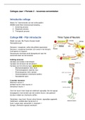
1. Electrical: If a neuron is active, it produces the action potentials. These action potentials do not
only lead to calcium entry to the cell (optical signal indirectly), but also just crunch. And because
crunch enters the cell, they generate an action potential. And thick metal sticks are sensitive to the
value of that potential and can indicate that.
2. Optical: Are invasive because you cannot use it if the skull is still there. You need to remove a part
of the skull. The imaging works by expressing fluorescent calcium indication inside the neurons,
which can then be lighted to photon the signal recorded. And the signal depends on for instance the
amount of calcium ions that are near the indicator. Because when the calcium indicator absorbs
calcium, it will indicate that it absorbs calcium.
3. Magnetic Resonance Molecular: not give you a very high resolution. It allows you to measure
indirect the activity of the neurons using the bold signal.
4. Molecular (= alternative methods):
So, we are interested in the spikes. And when a cell spikes it builds up for instance calcium
concentration. So, every time spikes happens than the Ca goes up. So, you get a signal inside the cell
(Intracellular activity indicator). You can bring in the cell DNA and say copy that DNA using a
polymerase. The polymerase will find the right piece and put it there, then it will proceed to the line
copying the whole piece of DNA. DNA polymerase can make mistakes so this will be checked and
removed.
→ The number of mistakes the polymerase makes, depends on concentrations in the cell. Depending
on the Ca concentration different number of mistakes are made. As this progress over time.
➔ As progresses over time you can see a record of mistakes arising over time, hence a record of the
intracellular indicator of the cell, hence indication of Ca concentration, hence indication of which
spikes were produced over time. At the end sequence it and reconstruct the history of each neuron.
Where do we measure neural
activity?
You can insert the electrodes into the brain and they are sensitive to a signal (extracellular potential
which reflects the activity of neurons). But there are more places where you can put electrodes. The
charactered of the signal changes if you go from outside to inside electrodes. The signal has to be
3
, really big outside, has to reflect the signal of many neurons. As you go down, the signal becomes more
sensitive for smaller groups of neurons.
EEG electrodes: on the skull you can put EEG electrodes.
Epidermal ECoG array: you can put this under the skull.
Subdural ECoG array: below the dura.
Intracortical microelectrode: below the arachnoid and put the electrode inside the grey matter.
Electrodes that go into the grey matter coming in many different shapes
and sizes.
Shank: you can measure on multiple location in the brain close by the signal. You can use different
locations in the cortex.
Utah: you can measure in different depths inside the cortex.
Cone: in vitro.
A couple of years ago, a new area started. One of these is called neuropixels. These are relatively large
electrodes. Shank goes into the brain (zoomed in in picture A).
You can record on each of them. 960 of these sides on the probe. You can pick which sides you are
most interested in, and 384 sides you found most interesting.
There are a couples of things that you want to
check:
- What is the impendence of the side you record
from? 50-250.
- What is the noise that is recorded? (4-8) Then you
know how big is should be in order be detectable
above the noise
- Because it is quite long, it goes through a lot of
different cortical and subcortical areas. (ab = each
of the little dot is a spike). → recordings are
impressive.
Concepts potential, current & resistance.
Current: circuit where we have battery and a lamp. And in this circuit current flows. The current is
produced by the battery and are positive / negative charges. That moves through the lamp, loose their
energy and then the current returns to the battery. The battery has plus and minus side. Voltage is the
4
Table of Contents
Lecture 2 Electrophysiology – electrical potentials in the brain (extra/intracellular, AD
range etc.) ........................................................................................................................ 9
1. Basis of membrane potential (sodium-potassium pump, Na, K, Cl). ........................................ 9
1. Extracellular recording ............................................................................................................................. 11
2. Intracellular recording ............................................................................................................................. 11
3. Patch clamp: ............................................................................................................................................ 11
Lecture 3 Structural and functional imaging .................................................................... 16
1. Two-photon imaging................................................................................................................................ 16
2. Structural imaging ................................................................................................................................... 20
3. Functional imaging .................................................................................................................................. 20
4. Two-photon imaging Applied in Visual neuroscience.............................................................................. 24
5. Combination two-photon imaging and other techniques. ...................................................................... 26
6. Future perspectives ................................................................................................................................. 26
Lecture 5 Optogenetics ................................................................................................... 27
1. Light sensitive channels. .......................................................................................................................... 27
2. Targeting: getting the channel where it needs to be. ............................................................................. 29
3. Light Delivery: getting the light where it needs to be. (Yizhar Neuron 2011) ......................................... 30
Channel models, with light induced switches. ............................................................................................ 31
4. Test light intensity of the fiber in order to make it fire ........................................................................... 31
5. Applications of optogenetics: .................................................................................................................. 32
Lecture 6 Magnetothermal stimulation ........................................................................... 37
Lecture 7 Genetic Methods .............................................................................................. 45
1. Gene delivery methods: Physical, chemical methods (transfection) ...................................................... 45
Viral methods .............................................................................................................................................. 46
1
,References:
Ba W, Selten MM, van der Raadt J, van Veen H
Busche, Science 2008
Chen et al., 2013 Nature
Chen et al., 2015
Christiansen et al., 2017.
Fenno, L., Yizhar, O. & Deisseroth, K. (2011) The development and application of optogenetics.
Annual review of neuroscience, 34, 389-412.
Foutz, T.J., Arlow, R.L. & McIntyre, C.C. (2012) Theoretical principles underlying optical stimulation
of a channelrhodopsin-2 positive pyramidal neuron. Journal of neurophysiology, 107, 3235-3245.
Friedl et al. (2007)
Hallet, 2007
Holtmaat and Svoboda, 2009
Jia et al., Nature 2010
James H. Marshel, Yoon Seok Kim, Timothy A. Machado, et. al. Cortical layer-specific critical
dynamics triggering perception (2019)
Krienberger and Konnerth, 2012)
Munshi et al (2017)
Ohki et al. (2006)
Ramirez. Steve, Xu Liu, et. al, (2013)
Reijmers et al, (2007)
Tominaga, 1998
Weijan Yang, Luis Carillo-Reid, Yuki Bando, et. al), Simultaneous two-photon imaging and two-
photon optogenetics of cortical circuits in three dimensions. (2018)
Zhang, F., Wang, L.P., Brauner, M., Liewald, J.F., Kay, K., Watzke, N., Wood, P.G., Bamberg, E.,
Nagel, G., Gottschalk, A. & Deisseroth, K. Multimodal fast optical interrogation of neural circuitry.
Nature, 446, 633-639. (2007)
2
,Abstract Methods in Neuroscience
Lecture 1 Electrodes.
Why do we use electrodes?
Interested in the brain → interested in the activity of the neurons in the brain. We can measure that
activity in number of different ways: for example, with electrodes.
1. Electrical: If a neuron is active, it produces the action potentials. These action potentials do not
only lead to calcium entry to the cell (optical signal indirectly), but also just crunch. And because
crunch enters the cell, they generate an action potential. And thick metal sticks are sensitive to the
value of that potential and can indicate that.
2. Optical: Are invasive because you cannot use it if the skull is still there. You need to remove a part
of the skull. The imaging works by expressing fluorescent calcium indication inside the neurons,
which can then be lighted to photon the signal recorded. And the signal depends on for instance the
amount of calcium ions that are near the indicator. Because when the calcium indicator absorbs
calcium, it will indicate that it absorbs calcium.
3. Magnetic Resonance Molecular: not give you a very high resolution. It allows you to measure
indirect the activity of the neurons using the bold signal.
4. Molecular (= alternative methods):
So, we are interested in the spikes. And when a cell spikes it builds up for instance calcium
concentration. So, every time spikes happens than the Ca goes up. So, you get a signal inside the cell
(Intracellular activity indicator). You can bring in the cell DNA and say copy that DNA using a
polymerase. The polymerase will find the right piece and put it there, then it will proceed to the line
copying the whole piece of DNA. DNA polymerase can make mistakes so this will be checked and
removed.
→ The number of mistakes the polymerase makes, depends on concentrations in the cell. Depending
on the Ca concentration different number of mistakes are made. As this progress over time.
➔ As progresses over time you can see a record of mistakes arising over time, hence a record of the
intracellular indicator of the cell, hence indication of Ca concentration, hence indication of which
spikes were produced over time. At the end sequence it and reconstruct the history of each neuron.
Where do we measure neural
activity?
You can insert the electrodes into the brain and they are sensitive to a signal (extracellular potential
which reflects the activity of neurons). But there are more places where you can put electrodes. The
charactered of the signal changes if you go from outside to inside electrodes. The signal has to be
3
, really big outside, has to reflect the signal of many neurons. As you go down, the signal becomes more
sensitive for smaller groups of neurons.
EEG electrodes: on the skull you can put EEG electrodes.
Epidermal ECoG array: you can put this under the skull.
Subdural ECoG array: below the dura.
Intracortical microelectrode: below the arachnoid and put the electrode inside the grey matter.
Electrodes that go into the grey matter coming in many different shapes
and sizes.
Shank: you can measure on multiple location in the brain close by the signal. You can use different
locations in the cortex.
Utah: you can measure in different depths inside the cortex.
Cone: in vitro.
A couple of years ago, a new area started. One of these is called neuropixels. These are relatively large
electrodes. Shank goes into the brain (zoomed in in picture A).
You can record on each of them. 960 of these sides on the probe. You can pick which sides you are
most interested in, and 384 sides you found most interesting.
There are a couples of things that you want to
check:
- What is the impendence of the side you record
from? 50-250.
- What is the noise that is recorded? (4-8) Then you
know how big is should be in order be detectable
above the noise
- Because it is quite long, it goes through a lot of
different cortical and subcortical areas. (ab = each
of the little dot is a spike). → recordings are
impressive.
Concepts potential, current & resistance.
Current: circuit where we have battery and a lamp. And in this circuit current flows. The current is
produced by the battery and are positive / negative charges. That moves through the lamp, loose their
energy and then the current returns to the battery. The battery has plus and minus side. Voltage is the
4