

COLLES’ FRACTURE
DEFINITION
This is a fracture at the distal end of the radius with dislocation of the inferior radio-ulnar joint, at its cortico-
cancellous junction about1.5 Inch or 2.5 cm from the distal articular surface and with typical displacement
in adults.
It is the commonest fracture in people above forty years of age, and is particularly common in women
because of postmenopausal osteoporosis.
MECHANISM OF INJURY
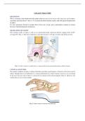
The common mode of injury is fall on an outstretched hand with dorsi-flexion ranging from 40-90°
(average 60°) (Fig. 1).The force required to cause this fracture is 192 kg in women and 282 kg in men.
Fig. 1: Colles’ fracture is usually due to a slip and fall on the outstretched hands in elderly females
CLINICAL FEATURES
The patient complains of pain, swelling, deformity and other usual features of fracture at the lower end of
radius. Though dinner fork deformity is a classical deformity in a Colles’fracture, however, it is not found
in all cases but seen only if there is a dorsal tilt or rotation of the distal fragment (Figs 2). However, the
styloid process test is more reliable.
Fig. 2: Colles’ fracture (A dinner fork deformity)
, Styloid Process Test
Normally, the radial styloid process is lower by 1.3 cm when compared to the ulnar styloid process. In
Colles’ fracture both radial and ulnar styloid processes are at the same level and are found in all
displacements of Colles’ fracture. Hence, this is a more reliable sign than dinner fork deformity (Figs 3 A
and B).
Figs 3 A and B: Styloid process test: (A) Normal (B) In Colles’ fractures
RADIOLOGY
Radiographs of the wrist (Figs 4 A and B) both AP and lateral views of the affected wrist and lower end
of the radius are taken. The points noted in the AP view are metaphyseal comminution, fracture line
extending into the radiocarpal or inferior radioulnar joint and fracture of the ulnar styloid process (seen
in about 60% of the cases). In the lateral view, the points noted are dorsal displacement and dorsal tilt
of the distal fragment, sharp palmar surface and dorsal comminution of the lower end of radius, distal
radioulnar joint subluxation, etc.
Figs 4 A and B: Radiographs showing Colles’ fracture: (A) AP view, and (B) lateral view
RELEVANT ANATOMY
The distal end of the radius articulates with the carpal bones (radio-carpal joint), and the distal end
of the ulna (radio-ulnar joint). Normally, the distal articular surface of the radius faces ventrally and
medially (Fig- 6). The tip of the radial styloid is about 1 cm distal to the tip of the ulnar styloid.
PATHOANATOMY
Displacement: The fracture line runs transversely at the cortico-cancellous junction. In the majority of
DEFINITION
This is a fracture at the distal end of the radius with dislocation of the inferior radio-ulnar joint, at its cortico-
cancellous junction about1.5 Inch or 2.5 cm from the distal articular surface and with typical displacement
in adults.
It is the commonest fracture in people above forty years of age, and is particularly common in women
because of postmenopausal osteoporosis.
MECHANISM OF INJURY
The common mode of injury is fall on an outstretched hand with dorsi-flexion ranging from 40-90°
(average 60°) (Fig. 1).The force required to cause this fracture is 192 kg in women and 282 kg in men.
Fig. 1: Colles’ fracture is usually due to a slip and fall on the outstretched hands in elderly females
CLINICAL FEATURES
The patient complains of pain, swelling, deformity and other usual features of fracture at the lower end of
radius. Though dinner fork deformity is a classical deformity in a Colles’fracture, however, it is not found
in all cases but seen only if there is a dorsal tilt or rotation of the distal fragment (Figs 2). However, the
styloid process test is more reliable.
Fig. 2: Colles’ fracture (A dinner fork deformity)
, Styloid Process Test
Normally, the radial styloid process is lower by 1.3 cm when compared to the ulnar styloid process. In
Colles’ fracture both radial and ulnar styloid processes are at the same level and are found in all
displacements of Colles’ fracture. Hence, this is a more reliable sign than dinner fork deformity (Figs 3 A
and B).
Figs 3 A and B: Styloid process test: (A) Normal (B) In Colles’ fractures
RADIOLOGY
Radiographs of the wrist (Figs 4 A and B) both AP and lateral views of the affected wrist and lower end
of the radius are taken. The points noted in the AP view are metaphyseal comminution, fracture line
extending into the radiocarpal or inferior radioulnar joint and fracture of the ulnar styloid process (seen
in about 60% of the cases). In the lateral view, the points noted are dorsal displacement and dorsal tilt
of the distal fragment, sharp palmar surface and dorsal comminution of the lower end of radius, distal
radioulnar joint subluxation, etc.
Figs 4 A and B: Radiographs showing Colles’ fracture: (A) AP view, and (B) lateral view
RELEVANT ANATOMY
The distal end of the radius articulates with the carpal bones (radio-carpal joint), and the distal end
of the ulna (radio-ulnar joint). Normally, the distal articular surface of the radius faces ventrally and
medially (Fig- 6). The tip of the radial styloid is about 1 cm distal to the tip of the ulnar styloid.
PATHOANATOMY
Displacement: The fracture line runs transversely at the cortico-cancellous junction. In the majority of

