Shad Ahmad Biology
Module 2 – Foundations in Biology
1 – Cell Structure
1) Light, Laser and Electron (TEM, SEM) Microscopes.
Light microscope:
• Use light. Coloured images produced.
• Have a low resolution and magnification.
• Images only 2D.
Laser Scanning Confocal Microscopes:
• Use laser beams to scan a specimen, which is usually tagged with a fluorescent dye.
• Laser causes dye to fluoresce (give off light). This light is then focused through a pinhole
onto a detector. The detector is hooked up to a computer, which generates a 2D and/or 3D
image.
• Pinhole blocks out-of-focus light, producing a much clearer image.
• Used to look at objects at different depths in thick specimens.
Electron Microscopes:
Use electrons to form an image. Higher resolution and magnification.
Transmission Electron Microscope (TEM):
• Use electromagnets to focus a beam of electrons through the specimen.
• Denser parts of the specimen absorb more electrons, making them look darker in the final
image.
• Higher resolution and magnification than SEMs.
• Only used on thin specimens.
• Only 2D images.
Scanning Electron Microscope (SEM):
• Scan a beam of electrons across the specimen. This knocks electrons from the specimen,
which are gathered in a cathode ray tube, to form an image.
• The images produced show the surface of the specimen.
• 2D and 3D images.
• Lower resolution and magnification than
TEMs.
,Shad Ahmad Biology
2) The preparation of microscope slides in light microscopy.
Dry Mount:
• To view a specimen under a light microscope, you need to stick it on a slide first.
• Your specimen must let light through to be seen clearly. Therefore, if you have a thick
specimen, you need to take a thin slice to use on your slide.
• Use tweezers to pick up the specimen.
• Put a cover slip on top.
Wet Mount:
• Put a small drop of water onto the slide.
• Use tweezers to place the specimen on top of the drop.
• Put the cover slip on upright, then carefully tilting and lowering it so it covers the specimen.
Try not to get any air bubbles there too.
• Once the cover slip is in position, add a stain. Put a drop of stain next to one edge of the
cover slip. Then put a paper towel next to the opposite edge. The stain gets drawn under the
slip, across the specimen.
3) How to use a light microscope.
1. Clip the slide with the specimen onto the stage.
2. Select the lowest magnification objective lens (4x, 10x, 40x)
3. Look down the eyepiece.
4. Use the coarse adjustment knob to raise and lower the stage to roughly focus.
5. Adjust the focus with the fine adjustment knob to get a clear image.
4) What is an eyepiece graticule and a stage micrometre?
• An eyepiece graticule is fitted onto the eyepiece. Like a ruler with numbers, but no units.
• Stage micrometre is placed on the stage – microscope slide with an accurate scale, and is
used to work out the value of the divisions on the eyepiece graticule at a particular
magnification, so you can replace the stage micrometre with the slide of the specimen and
measure its size.
,Shad Ahmad Biology
5) The use of staining in light and electron microscopy.
Staining samples for light microscopes:
• Staining is done by dye, EG: methylene blue, eosin.
• The stain is taken up by some parts of the objects more than others, leading to a contrast,
which makes the different parts show up.
• Different stains are used to make different things show up. EG: eosin used to stain cell
cytoplasm; methylene blue stains DNA.
Staining samples for electron microscopes:
• Objects are dipped in a solution of heavy metals ions.
• The metal ions scatter the electrons, creating contrast.
6) What is the equation for magnification?
Total magnification = Eyepiece lens magnification x Objective lens magnification
Milli = 10-3 m
Micro = 10-6 m
Nano = 10-9 m
Pico = 10-12 m
7) What is the difference between magnification and resolution?
Magnification = How many times the image is enlarged from the sample.
Resolution = How detailed the image is, and how well a microscope distinguishes between two
points that are close together.
,Shad Ahmad Biology
8) The ultrastructure of eukaryotic cells and the functions of the different cellular components.
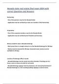
(phospholipid bilayer)
- Prevents plant cells from bursting when turgid
- Maintains cell’s shape
- They are permeable and allow solutions to pass through
- Fungi have cell walls that contain chitin, not cellulose
Nucleoplasm
Chromatin = DNA wrapped
around a histone protein.
- Ribosomes bound to the exterior of RER
rRNA
are mainly for synthesizing proteins that
will be exported outside the cell
- Ribosomes that are free in the cytoplasm,
Protein assemble proteins that will be used inside
the cell
,Shad Ahmad Biology
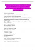
Cisternae = fluid-filled spaceç RER is the intracellular transport
system: the cisternae form channels
for transporting substances from one
area of a cell to another
SER contains enzymes that catalyse
Cisternae = fluid-filled space reactions involved with lipid metabolism,
such as the synthesis of cholesterol, lipids
and steroid hormones.
Proteins are modified
by e.g. adding sugar
molecules to make
glycoproteins, adding
lipid molecules to make
lipoproteins or being
folded into their 3D
Cisternae = fluid-filled space
shape.
They are self-replicating, so more can be
made if the cell’s energy needs to
Contains circular DNA. increase. They are abundant in cells where
much metabolic activity takes place.
Chloroplast
envelope
(outer +
inner
membrane
+ inter
membrane
space Contains circular DNA.
, Shad Ahmad Biology
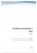
Microtubules are made of tubulin proteins
The only type of human cell to have an
undulipodium (a longer cilium) is a
sperm. The undulipodium enables the
sperm to move. In prokaryotic cells
they are called flagella.
Vacuole
The vacuole is surrounded by a membrane called the tonoplast, and contains fluid (cell sap).
It is filled with water and solutes and maintains cell stability, because when full it pushes against
the cell wall, making the cell turgid.
Thus, if all the plant cells are turgid then this helps to support the plant, especially in non-woody
plants.
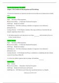
9) Eukaryotic Cell Structure (EG: animal and plant cells).
Module 2 – Foundations in Biology
1 – Cell Structure
1) Light, Laser and Electron (TEM, SEM) Microscopes.
Light microscope:
• Use light. Coloured images produced.
• Have a low resolution and magnification.
• Images only 2D.
Laser Scanning Confocal Microscopes:
• Use laser beams to scan a specimen, which is usually tagged with a fluorescent dye.
• Laser causes dye to fluoresce (give off light). This light is then focused through a pinhole
onto a detector. The detector is hooked up to a computer, which generates a 2D and/or 3D
image.
• Pinhole blocks out-of-focus light, producing a much clearer image.
• Used to look at objects at different depths in thick specimens.
Electron Microscopes:
Use electrons to form an image. Higher resolution and magnification.
Transmission Electron Microscope (TEM):
• Use electromagnets to focus a beam of electrons through the specimen.
• Denser parts of the specimen absorb more electrons, making them look darker in the final
image.
• Higher resolution and magnification than SEMs.
• Only used on thin specimens.
• Only 2D images.
Scanning Electron Microscope (SEM):
• Scan a beam of electrons across the specimen. This knocks electrons from the specimen,
which are gathered in a cathode ray tube, to form an image.
• The images produced show the surface of the specimen.
• 2D and 3D images.
• Lower resolution and magnification than
TEMs.
,Shad Ahmad Biology
2) The preparation of microscope slides in light microscopy.
Dry Mount:
• To view a specimen under a light microscope, you need to stick it on a slide first.
• Your specimen must let light through to be seen clearly. Therefore, if you have a thick
specimen, you need to take a thin slice to use on your slide.
• Use tweezers to pick up the specimen.
• Put a cover slip on top.
Wet Mount:
• Put a small drop of water onto the slide.
• Use tweezers to place the specimen on top of the drop.
• Put the cover slip on upright, then carefully tilting and lowering it so it covers the specimen.
Try not to get any air bubbles there too.
• Once the cover slip is in position, add a stain. Put a drop of stain next to one edge of the
cover slip. Then put a paper towel next to the opposite edge. The stain gets drawn under the
slip, across the specimen.
3) How to use a light microscope.
1. Clip the slide with the specimen onto the stage.
2. Select the lowest magnification objective lens (4x, 10x, 40x)
3. Look down the eyepiece.
4. Use the coarse adjustment knob to raise and lower the stage to roughly focus.
5. Adjust the focus with the fine adjustment knob to get a clear image.
4) What is an eyepiece graticule and a stage micrometre?
• An eyepiece graticule is fitted onto the eyepiece. Like a ruler with numbers, but no units.
• Stage micrometre is placed on the stage – microscope slide with an accurate scale, and is
used to work out the value of the divisions on the eyepiece graticule at a particular
magnification, so you can replace the stage micrometre with the slide of the specimen and
measure its size.
,Shad Ahmad Biology
5) The use of staining in light and electron microscopy.
Staining samples for light microscopes:
• Staining is done by dye, EG: methylene blue, eosin.
• The stain is taken up by some parts of the objects more than others, leading to a contrast,
which makes the different parts show up.
• Different stains are used to make different things show up. EG: eosin used to stain cell
cytoplasm; methylene blue stains DNA.
Staining samples for electron microscopes:
• Objects are dipped in a solution of heavy metals ions.
• The metal ions scatter the electrons, creating contrast.
6) What is the equation for magnification?
Total magnification = Eyepiece lens magnification x Objective lens magnification
Milli = 10-3 m
Micro = 10-6 m
Nano = 10-9 m
Pico = 10-12 m
7) What is the difference between magnification and resolution?
Magnification = How many times the image is enlarged from the sample.
Resolution = How detailed the image is, and how well a microscope distinguishes between two
points that are close together.
,Shad Ahmad Biology
8) The ultrastructure of eukaryotic cells and the functions of the different cellular components.
(phospholipid bilayer)
- Prevents plant cells from bursting when turgid
- Maintains cell’s shape
- They are permeable and allow solutions to pass through
- Fungi have cell walls that contain chitin, not cellulose
Nucleoplasm
Chromatin = DNA wrapped
around a histone protein.
- Ribosomes bound to the exterior of RER
rRNA
are mainly for synthesizing proteins that
will be exported outside the cell
- Ribosomes that are free in the cytoplasm,
Protein assemble proteins that will be used inside
the cell
,Shad Ahmad Biology
Cisternae = fluid-filled spaceç RER is the intracellular transport
system: the cisternae form channels
for transporting substances from one
area of a cell to another
SER contains enzymes that catalyse
Cisternae = fluid-filled space reactions involved with lipid metabolism,
such as the synthesis of cholesterol, lipids
and steroid hormones.
Proteins are modified
by e.g. adding sugar
molecules to make
glycoproteins, adding
lipid molecules to make
lipoproteins or being
folded into their 3D
Cisternae = fluid-filled space
shape.
They are self-replicating, so more can be
made if the cell’s energy needs to
Contains circular DNA. increase. They are abundant in cells where
much metabolic activity takes place.
Chloroplast
envelope
(outer +
inner
membrane
+ inter
membrane
space Contains circular DNA.
, Shad Ahmad Biology
Microtubules are made of tubulin proteins
The only type of human cell to have an
undulipodium (a longer cilium) is a
sperm. The undulipodium enables the
sperm to move. In prokaryotic cells
they are called flagella.
Vacuole
The vacuole is surrounded by a membrane called the tonoplast, and contains fluid (cell sap).
It is filled with water and solutes and maintains cell stability, because when full it pushes against
the cell wall, making the cell turgid.
Thus, if all the plant cells are turgid then this helps to support the plant, especially in non-woody
plants.
9) Eukaryotic Cell Structure (EG: animal and plant cells).