Moleculaire Biologie
H7 Cell Structure and Function
7.1 Biologists use microscopes and biochemistry to study cells
Microscopy
The microscopes first used by Renaissance scientists, as well as the microscopes you are likely to use
in the laboratory are all light microscopes. In a light microscope (LM), visible light is passed through
the specimen and then through glass lenses. The lenses refract the light in such a way that the
image of the specimen is magnified as it is projected into the eye or camera.
Three important parameters in microscopy are magnification, resolution and contrast.
- Magnification is the ratio of an object’s image size to its real size. Light microscopes can
magnify effectively to about 1,000 times the actual size of the specimen; at greater
magnifications, additional details cannot be seen clearly.
- Resolution is a measure of the clarity of the image; it is the minimum distance two points
can be separated and still be distinguished at separate points.
- Contrast is the difference between in brightness between the light and dark areas of an
image. Methods for enhancing contrast include staining or labelling cell components to
stand out visually.
Until recently, the resolution barrier prevented cell biologists from using standard light microscopy
when studying organelles ,the membrane-closed structures within eukaryotic cells. To view these
structures, the electron microscope (EM) was introduced. EM focuses a beam of electrons through
the specimen or onto its surface. Resolution is inversely related to the wavelength of the light a
microscope uses for imaging, and electron beams have much shorter wavelengths than visible light.
The scanning electron microscope (SEM) is especially useful for detailed study of the topography of
specimen. The electron beam scans the surface of the sample, usually coated with a film of gold. The
beam excites electrons on the surface, and these secondary electrons are detected by a device that
translates the pattern of electrons onto an electronic signal sent to a video screen. The transmission
electron microscope (TEM) is used to study the internal structure of cells. The TEM aims an electron
beam through a very thin section of the specimen, much as a light microscope aims light through a
sample on a slide. For the TEM, the specimen has been stained with atoms of heavy metals, which
attach to certain cellular structures, thus enhancing the electron diversity of some parts of the cell
more than others.
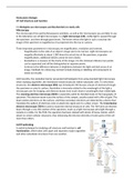
Cell Fractioning
A useful technique for studying cell structure and function is cell
fractionation, which takes cells apart and separates major organelles
and other subcellular structures from one another.
, 7.2 Eukaryotic cells have internal membranes that compartmentalize their functions
Cells – the basic structural and functional units if every organism – are of two distinct types:
prokaryotic and eukaryotic. Organisms of the domains Bacteria and Archaea consists of prokaryotic
cells. Organisms of the domain Eukarya – protists, fungi, animals, and plants – all consists of
eukaryotic cells.
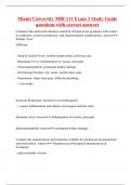
Comparing Prokaryotic and Eukaryotic Cells
All cells share certain basic features: They are all bounded by a selective barrier, called the plasma
membrane. Inside all cells is a semifluid, jellylike substance called the cytosol, in which subcellular
components are suspended. All cells contain chromosomes, which carry genes in the form of DNA.
And all cells have ribosomes, tiny complexes that make proteins according to instructions from the
genes.
- A major difference between prokaryotic and eukaryotic cells is the location of their DNA. In a
eukaryotic cell, most of the DNA is in an organelle called the nucleus, which is bounded by a
double membrane. In a prokaryotic cell, the DNA is concentrated in a region that is not
membrane-enclosed, called the nucleoid.
The interior of either type of cell is called the cytoplasm; in eukaryotic cells, this term refers only to
the region between the nucleus and the plasma membrane. Eukaryotic cells are generally much
larger than prokaryotic cells. Size is a general feature of cell structure that relates to function.
Metabolic requirements also impose theoretical upper limits on the size that is practical for a single
cell. At the boundary of every cell, the plasma membrane functions as a selective barrier that allows
passage of oxygen, nutrients, and wastes to service the entire cell. For each square micrometre of
membrane, only a limited amount of particular substance can cross per second, so the ratio of
surface area to volume is critical. As a cell increases in size, its surface area grows proportionately
less than its volume.
H7 Cell Structure and Function
7.1 Biologists use microscopes and biochemistry to study cells
Microscopy
The microscopes first used by Renaissance scientists, as well as the microscopes you are likely to use
in the laboratory are all light microscopes. In a light microscope (LM), visible light is passed through
the specimen and then through glass lenses. The lenses refract the light in such a way that the
image of the specimen is magnified as it is projected into the eye or camera.
Three important parameters in microscopy are magnification, resolution and contrast.
- Magnification is the ratio of an object’s image size to its real size. Light microscopes can
magnify effectively to about 1,000 times the actual size of the specimen; at greater
magnifications, additional details cannot be seen clearly.
- Resolution is a measure of the clarity of the image; it is the minimum distance two points
can be separated and still be distinguished at separate points.
- Contrast is the difference between in brightness between the light and dark areas of an
image. Methods for enhancing contrast include staining or labelling cell components to
stand out visually.
Until recently, the resolution barrier prevented cell biologists from using standard light microscopy
when studying organelles ,the membrane-closed structures within eukaryotic cells. To view these
structures, the electron microscope (EM) was introduced. EM focuses a beam of electrons through
the specimen or onto its surface. Resolution is inversely related to the wavelength of the light a
microscope uses for imaging, and electron beams have much shorter wavelengths than visible light.
The scanning electron microscope (SEM) is especially useful for detailed study of the topography of
specimen. The electron beam scans the surface of the sample, usually coated with a film of gold. The
beam excites electrons on the surface, and these secondary electrons are detected by a device that
translates the pattern of electrons onto an electronic signal sent to a video screen. The transmission
electron microscope (TEM) is used to study the internal structure of cells. The TEM aims an electron
beam through a very thin section of the specimen, much as a light microscope aims light through a
sample on a slide. For the TEM, the specimen has been stained with atoms of heavy metals, which
attach to certain cellular structures, thus enhancing the electron diversity of some parts of the cell
more than others.
Cell Fractioning
A useful technique for studying cell structure and function is cell
fractionation, which takes cells apart and separates major organelles
and other subcellular structures from one another.
, 7.2 Eukaryotic cells have internal membranes that compartmentalize their functions
Cells – the basic structural and functional units if every organism – are of two distinct types:
prokaryotic and eukaryotic. Organisms of the domains Bacteria and Archaea consists of prokaryotic
cells. Organisms of the domain Eukarya – protists, fungi, animals, and plants – all consists of
eukaryotic cells.
Comparing Prokaryotic and Eukaryotic Cells
All cells share certain basic features: They are all bounded by a selective barrier, called the plasma
membrane. Inside all cells is a semifluid, jellylike substance called the cytosol, in which subcellular
components are suspended. All cells contain chromosomes, which carry genes in the form of DNA.
And all cells have ribosomes, tiny complexes that make proteins according to instructions from the
genes.
- A major difference between prokaryotic and eukaryotic cells is the location of their DNA. In a
eukaryotic cell, most of the DNA is in an organelle called the nucleus, which is bounded by a
double membrane. In a prokaryotic cell, the DNA is concentrated in a region that is not
membrane-enclosed, called the nucleoid.
The interior of either type of cell is called the cytoplasm; in eukaryotic cells, this term refers only to
the region between the nucleus and the plasma membrane. Eukaryotic cells are generally much
larger than prokaryotic cells. Size is a general feature of cell structure that relates to function.
Metabolic requirements also impose theoretical upper limits on the size that is practical for a single
cell. At the boundary of every cell, the plasma membrane functions as a selective barrier that allows
passage of oxygen, nutrients, and wastes to service the entire cell. For each square micrometre of
membrane, only a limited amount of particular substance can cross per second, so the ratio of
surface area to volume is critical. As a cell increases in size, its surface area grows proportionately
less than its volume.