Problem 2 2.4
Connections
The visual system
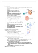
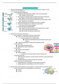
• Most neural signals from the retina travel out of the eye in the optic nerve to the
lateral geniculate nucleus (LGN) in the
thalamus
• Signals then travel to the primary visual
receiving area in the occipital lobe of the
cortex
• Striate cortex – the visual receiving area of the
cortex in which white stripes are created by
nerve fibres that run through it
• From here, signals are transmitted along two
pathways, one to the temporal lobe and the
other to the parietal lobe (blue arrows)
- Visual signals also reach areas in the frontal
lobe of the brain
• Superior colliculus – area involved in
controlling eye movements and other visual behaviours
that receives about 10% of the fibres from the optic
nerve
• Signals from half of each retina cross over to the
opposite side of the brain
• Lateral geniculate nucleus – first major area where
visual signals are received
Processing in the Lateral Geniculate Nucleus
Receptive fields of the LGN neurons
• LGN neurons have the same centre-surround
configuration as retinal ganglion cells
• They respond best to small spots of light on the retina
• Major function is to regulate and organise neural
information as it flows from the retina to the visual
cortex
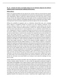
Information flow in the lateral geniculate nucleus
• 90% of the fibres in the optic nerve arrive at the LGN
- 10% travel to the superior colliculus
• The LGN also receives signals from the cortex, from the brain stem, from other
neurons in the thalamus (T) and from other neurons in the LGN (L)
- It then sends its output to the cortex
• LGN receives more input back from the cortex than it receives from the retina
- For every 10 nerve impulses it receives from the retina, it sends only 4 to the
cortex
• The smallest signal of all is from the LGN to the cortex
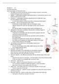
Organisation by left and right eyes
, • The LGN is a bilateral structure meaning there is one LGN
in the left hemisphere and one in the right hemisphere
• Each nucleus has 6 layers. Each layer receives signals
from only one eye
- Layers 2,3 and 5 (red) receive signals from the
ipsilateral eye (same side of body as the LGN)
- Layers 1,4 and 6 (blue) receive signals from the
contralateral eye (opposite side of body as LGN)
• each eye sends half of its neurons to the LGN that is
located in the left hemisphere and half to the LGN in the
right hemisphere
• because the signals from each eye are sorted into
different layers, the information from the left and right
eyes is kept separated in the LGN
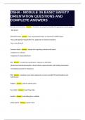
organisation as a spatial map
• when the man looks at the cup points A, B and C on the
cup are imaged on points A, B and C of the retina
• each place on the retina corresponds to a specific place
on the LGN
• retinotopic map – a map in which each point on the LGN corresponds to a point on
the retina
- occur in each of the other layers as well, and the
maps of each layer line up with one another
• this correspondence means that neurons entering the
LGN are arranged so that fibres carrying signals from
the same area of the retina end up in the same area of
the LGN
- thus, the receptive fields of neurons that are near
each other in the LGN are adjacent to each other
at the respective points on the retina
• if we were to lower an electrode perpendicularly, all
the neurons we encounter along the electrode track
will have receptive fields at the same location on the
retina
- 1 mil ganglion cell fibres travel to each LGN and then travel to the correct LGN
layer, as well as finding its way to a location next to other fibres that left from
the same place on the retina
• The result is aligned, overlapping retinotopic maps in each of the LGN’s six layers
Receptive Fields of Neurons in the striate cortex
• More than 80% of the cortex responds to visual stimuli
- Most of the cortex responds when the retina is stimulated
• Hubel and Wiesel
- Found cells in the striate cortex with receptive fields that, like centre-surround
receptive fields of neurons in the retina and the LGN, have excitatory and
inhibitory areas
, - these areas are arranged side by side rather than in the centre-surround
configuration
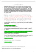
• simple cortical cells – these cells with side-by-side receptive fields
- a cell with this receptive field would respond best to vertical bars
• a vertical bar that illuminates only the excitatory areas causes high firing, but as the
bar is tilted so the inhibitory area is illuminated, firing decreases
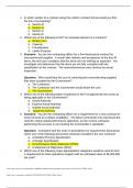
• the relationship between orientation and firing is indicated by neurons orientation
tuning curve - this is determined by measuring the response of a simple cortical cell
to bars with different orientations
• The tuning curve in the third image shows that the cell responds with 25 nerve
impulses per second to a vertically orientated bar and that the cells response
decreases as the bar is tilted away from the vertical, and begins stimulating
inhibitory areas of the neurons receptive field
• Many cortical neurons respond best to moving bar-like stimuli with specific
orientations
• complex cells, like simple cells, respond
best to bars of a particular orientation.
• unlike simple cells, which respond to
small spots of light or to stationary
stimuli, most complex cells respond
only when a correctly orientated power
of light moves across the entire
receptive field
• many complex cells respond best to a
particular direction of movement
• Because these neurons don't respond to stationary flashes of light, their receptive
fields are not indicated buy pluses or minuses, but by indicating the area which,
when stimulated, elicits a response in the neuron
• End-stopped cells - another type of cell that fire to moving lines of a specific link or
two moving corners or angles
- The neurons response increases as the corner shape stimulus gets longer but
then stops responding when that corner becomes too long
Connections
The visual system
• Most neural signals from the retina travel out of the eye in the optic nerve to the
lateral geniculate nucleus (LGN) in the
thalamus
• Signals then travel to the primary visual
receiving area in the occipital lobe of the
cortex
• Striate cortex – the visual receiving area of the
cortex in which white stripes are created by
nerve fibres that run through it
• From here, signals are transmitted along two
pathways, one to the temporal lobe and the
other to the parietal lobe (blue arrows)
- Visual signals also reach areas in the frontal
lobe of the brain
• Superior colliculus – area involved in
controlling eye movements and other visual behaviours
that receives about 10% of the fibres from the optic
nerve
• Signals from half of each retina cross over to the
opposite side of the brain
• Lateral geniculate nucleus – first major area where
visual signals are received
Processing in the Lateral Geniculate Nucleus
Receptive fields of the LGN neurons
• LGN neurons have the same centre-surround
configuration as retinal ganglion cells
• They respond best to small spots of light on the retina
• Major function is to regulate and organise neural
information as it flows from the retina to the visual
cortex
Information flow in the lateral geniculate nucleus
• 90% of the fibres in the optic nerve arrive at the LGN
- 10% travel to the superior colliculus
• The LGN also receives signals from the cortex, from the brain stem, from other
neurons in the thalamus (T) and from other neurons in the LGN (L)
- It then sends its output to the cortex
• LGN receives more input back from the cortex than it receives from the retina
- For every 10 nerve impulses it receives from the retina, it sends only 4 to the
cortex
• The smallest signal of all is from the LGN to the cortex
Organisation by left and right eyes
, • The LGN is a bilateral structure meaning there is one LGN
in the left hemisphere and one in the right hemisphere
• Each nucleus has 6 layers. Each layer receives signals
from only one eye
- Layers 2,3 and 5 (red) receive signals from the
ipsilateral eye (same side of body as the LGN)
- Layers 1,4 and 6 (blue) receive signals from the
contralateral eye (opposite side of body as LGN)
• each eye sends half of its neurons to the LGN that is
located in the left hemisphere and half to the LGN in the
right hemisphere
• because the signals from each eye are sorted into
different layers, the information from the left and right
eyes is kept separated in the LGN
organisation as a spatial map
• when the man looks at the cup points A, B and C on the
cup are imaged on points A, B and C of the retina
• each place on the retina corresponds to a specific place
on the LGN
• retinotopic map – a map in which each point on the LGN corresponds to a point on
the retina
- occur in each of the other layers as well, and the
maps of each layer line up with one another
• this correspondence means that neurons entering the
LGN are arranged so that fibres carrying signals from
the same area of the retina end up in the same area of
the LGN
- thus, the receptive fields of neurons that are near
each other in the LGN are adjacent to each other
at the respective points on the retina
• if we were to lower an electrode perpendicularly, all
the neurons we encounter along the electrode track
will have receptive fields at the same location on the
retina
- 1 mil ganglion cell fibres travel to each LGN and then travel to the correct LGN
layer, as well as finding its way to a location next to other fibres that left from
the same place on the retina
• The result is aligned, overlapping retinotopic maps in each of the LGN’s six layers
Receptive Fields of Neurons in the striate cortex
• More than 80% of the cortex responds to visual stimuli
- Most of the cortex responds when the retina is stimulated
• Hubel and Wiesel
- Found cells in the striate cortex with receptive fields that, like centre-surround
receptive fields of neurons in the retina and the LGN, have excitatory and
inhibitory areas
, - these areas are arranged side by side rather than in the centre-surround
configuration
• simple cortical cells – these cells with side-by-side receptive fields
- a cell with this receptive field would respond best to vertical bars
• a vertical bar that illuminates only the excitatory areas causes high firing, but as the
bar is tilted so the inhibitory area is illuminated, firing decreases
• the relationship between orientation and firing is indicated by neurons orientation
tuning curve - this is determined by measuring the response of a simple cortical cell
to bars with different orientations
• The tuning curve in the third image shows that the cell responds with 25 nerve
impulses per second to a vertically orientated bar and that the cells response
decreases as the bar is tilted away from the vertical, and begins stimulating
inhibitory areas of the neurons receptive field
• Many cortical neurons respond best to moving bar-like stimuli with specific
orientations
• complex cells, like simple cells, respond
best to bars of a particular orientation.
• unlike simple cells, which respond to
small spots of light or to stationary
stimuli, most complex cells respond
only when a correctly orientated power
of light moves across the entire
receptive field
• many complex cells respond best to a
particular direction of movement
• Because these neurons don't respond to stationary flashes of light, their receptive
fields are not indicated buy pluses or minuses, but by indicating the area which,
when stimulated, elicits a response in the neuron
• End-stopped cells - another type of cell that fire to moving lines of a specific link or
two moving corners or angles
- The neurons response increases as the corner shape stimulus gets longer but
then stops responding when that corner becomes too long