NU 545
Unit 2 Study Guide
Advanced Pathophysiology
University of South Alabama.
This document provides a focused
study guide
It summarizes key concepts, lecture highlights, and
exam-relevant material to support efficient last-minute
review. The guide is structured to help students
reinforce understanding, identify weak areas, and prepare
confidently for the assessment.
, Study Guide Unit 2
Pathologic Alterations: Organs and Systems
Afferent Afferent means towards
Efferent means away from.
vs. In this instance, it means either toward or away
from the spine.
Efferent
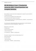
Cerebral
vs.
Cerebellar
CHAPTER 15
Key words:
What nerves are capable of regenerating?
(eBook key search term: “limited to myelinated fibers”)
Mature neurons do not divide and injury in the CNS causes permanent loss of damaged neurons. Crushed nerves recover better than cut
nerves. Peripheral nerves can repair themselves through axonal reaction
Local changes occur when the axon is severed
1. The cut ends retract and the axolemma covers the cut ends, diminishing the escape of axoplasm
2. Macrophages and Schwann cells begin to phagocytize damaged tissue
3. The cell body undergoes chromotolysis with swelling, loss of Nissl bodies, and the lateral migration of the nucleus
4. Antegrade (Wallerian) degeneration occurs in the distal axon
5. A characteristic swelling appears in the axon terminal and it degenerates and loses contact with the post synaptic membrane
within 7 days
6. Macrophages and Schwann cells phagocytize the remnants of the axon terminal
7. Schwann cells proliferate, forming a column or tube of Schwann cells enclosed by the original basal lamina of the
endoneurium.
8. Retrograde changes occur at the proximal end of the injured axon and are similar to antegrade changes but only back to the
next node of Ranvier.
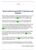
Approximately 7-14 days after the injury, new terminal sprouts project from the proximal segment guided by Schwann cells and
enter the sustaining substrate of a more detailed representation of these events
o This process is very slow, about 1mm/day, and is limited to myelinated fibers in the PNS.
o The closer the injury is to the cell body of the nerve, the greater the chances that the nerve cell will die and not regenerate
, o Peripheral nerves injured close to the spinal cord recover poorly and slowly because of the long distance between the
cell body and the peripheral termination of the axon.
The regeneration of axonal constituents in the CNS is limited by an increased incidence of glial scar formation (gliosis) and the
different nature of myelin formed by the oligodendrocyte
Nerve regeneration depends on many factors:
1. Location of the injury
2. The type of injury: crushing injury allows recovery more fully than does a cut injury
a. Crushed nerves sometimes fully recover, whereas cut nerves often form connective tissue scars that block or slow
regenerating axonal branches
3. The presence of inflammatory responses
4. The process of scarring
Review the anatomy of the brain.
(eBook key search term: “receives 15%”)
The Brain: allows individuals to reason, function intellectually, express personality and mood, and interact with the environment.
Weighs approximately 3 lbs. but receives 15%-20% of total cardiac output
THREE MAJOR DIVISIONS OF THE BRAIN
Structural The Reticular
Divisions of the Activating
Brain System
PRIMARY VESICLES SECONDARY VESICLES ASSOCIATED STRUCTURES
FOREBRAIN (prosencephalon) Cerebral hemispheres
Telencephalon Cerebral cortex
Rhinencephalon (olfaction)
Basal ganglia
Epithalamus
Diencephalon Thalamus
Hypothalamus
Subthalamus
MIDBRAIN Mesencephalon Tectum (corpora quadrigemina)
Connects the Pons to the diencephalon Tegmentum
Red nucleus
Substantia nigra
Cerebral peduncles
HINDBRAIN (rhombencephalon) Mentencephalon Cerebellum
Pons
Myelencephalon Medulla Oblongata
BRAINSTEM Comprised of the midbrain, pons, and
medulla oblongata
Connects the left / right hemispheres,
cerebellum, and spinal cord
Reticular formation / reticular activating
system
SPINAL CORD Spinal Cord
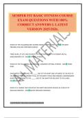
, CEREBRAL HEMISPHERES
Functional areas
of the cerebral
cortex
Left Hemisphere or cerebrum (lateral view) (midsagittal
view)
Functional areas
of the cerebral
cortex (lateral
view)
Cerebellum
(posterior view);
coordination of
voluntary
movement,
balance, and
posture
Unit 2 Study Guide
Advanced Pathophysiology
University of South Alabama.
This document provides a focused
study guide
It summarizes key concepts, lecture highlights, and
exam-relevant material to support efficient last-minute
review. The guide is structured to help students
reinforce understanding, identify weak areas, and prepare
confidently for the assessment.
, Study Guide Unit 2
Pathologic Alterations: Organs and Systems
Afferent Afferent means towards
Efferent means away from.
vs. In this instance, it means either toward or away
from the spine.
Efferent
Cerebral
vs.
Cerebellar
CHAPTER 15
Key words:
What nerves are capable of regenerating?
(eBook key search term: “limited to myelinated fibers”)
Mature neurons do not divide and injury in the CNS causes permanent loss of damaged neurons. Crushed nerves recover better than cut
nerves. Peripheral nerves can repair themselves through axonal reaction
Local changes occur when the axon is severed
1. The cut ends retract and the axolemma covers the cut ends, diminishing the escape of axoplasm
2. Macrophages and Schwann cells begin to phagocytize damaged tissue
3. The cell body undergoes chromotolysis with swelling, loss of Nissl bodies, and the lateral migration of the nucleus
4. Antegrade (Wallerian) degeneration occurs in the distal axon
5. A characteristic swelling appears in the axon terminal and it degenerates and loses contact with the post synaptic membrane
within 7 days
6. Macrophages and Schwann cells phagocytize the remnants of the axon terminal
7. Schwann cells proliferate, forming a column or tube of Schwann cells enclosed by the original basal lamina of the
endoneurium.
8. Retrograde changes occur at the proximal end of the injured axon and are similar to antegrade changes but only back to the
next node of Ranvier.
Approximately 7-14 days after the injury, new terminal sprouts project from the proximal segment guided by Schwann cells and
enter the sustaining substrate of a more detailed representation of these events
o This process is very slow, about 1mm/day, and is limited to myelinated fibers in the PNS.
o The closer the injury is to the cell body of the nerve, the greater the chances that the nerve cell will die and not regenerate
, o Peripheral nerves injured close to the spinal cord recover poorly and slowly because of the long distance between the
cell body and the peripheral termination of the axon.
The regeneration of axonal constituents in the CNS is limited by an increased incidence of glial scar formation (gliosis) and the
different nature of myelin formed by the oligodendrocyte
Nerve regeneration depends on many factors:
1. Location of the injury
2. The type of injury: crushing injury allows recovery more fully than does a cut injury
a. Crushed nerves sometimes fully recover, whereas cut nerves often form connective tissue scars that block or slow
regenerating axonal branches
3. The presence of inflammatory responses
4. The process of scarring
Review the anatomy of the brain.
(eBook key search term: “receives 15%”)
The Brain: allows individuals to reason, function intellectually, express personality and mood, and interact with the environment.
Weighs approximately 3 lbs. but receives 15%-20% of total cardiac output
THREE MAJOR DIVISIONS OF THE BRAIN
Structural The Reticular
Divisions of the Activating
Brain System
PRIMARY VESICLES SECONDARY VESICLES ASSOCIATED STRUCTURES
FOREBRAIN (prosencephalon) Cerebral hemispheres
Telencephalon Cerebral cortex
Rhinencephalon (olfaction)
Basal ganglia
Epithalamus
Diencephalon Thalamus
Hypothalamus
Subthalamus
MIDBRAIN Mesencephalon Tectum (corpora quadrigemina)
Connects the Pons to the diencephalon Tegmentum
Red nucleus
Substantia nigra
Cerebral peduncles
HINDBRAIN (rhombencephalon) Mentencephalon Cerebellum
Pons
Myelencephalon Medulla Oblongata
BRAINSTEM Comprised of the midbrain, pons, and
medulla oblongata
Connects the left / right hemispheres,
cerebellum, and spinal cord
Reticular formation / reticular activating
system
SPINAL CORD Spinal Cord
, CEREBRAL HEMISPHERES
Functional areas
of the cerebral
cortex
Left Hemisphere or cerebrum (lateral view) (midsagittal
view)
Functional areas
of the cerebral
cortex (lateral
view)
Cerebellum
(posterior view);
coordination of
voluntary
movement,
balance, and
posture