BIO 171 Module 3-6 Study Guide
Module Three: Microscopy
Microscopy: Using light or electrons to magnify objects.
History Behind Microscopy:
- Dating back to the 1600’s, glass lenses have been used to enlarge or magnify objects of
interest.
- The microscope is perhaps the most important resource for studying biology at a
microscopic level.
Units of Measurement:
- Micrometer: equals 10-6 m, one millionth of a meter
- Nanometer: equals 10-9 m, one billionth of a meter
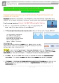
Factors that influence our ability to see an object:
- Resolution: the distance between two objects at which the objects can still be seen as
separate.
o The closer the two objects are to each other, the higher the resolution
requirement will be to maintain viewing the two objects as separate.
- Contrast: The difference in light absorbance between two areas or objects.
o The lower the contract between an object and its background, the harder it will
be to see that object.
o The greater the contrast between two areas will make it easier to see
- Magnification: Increase in the size of the object. Results when light bends as it passes
through a lens.
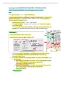
Staining Microbes:
- Staining is using dyes to improve and increases the resolution and contrast of a sample
for visualization via light microscopy (when
trying to see them in the microscopes.).
- The different types of stains explain several
different things such as classifying and
identifying microbes based
- Benefits of Staining: on their traits.
o Increase contrast o Allows for
determining cellular…
Shape
Size
Arrangement
Number of cells in sample Preparing Specimens
for Staining:
- Samples must be prepped for staining
o Steps for preparing a slide:
Smear or Place Specimen on Slide
• Liquid Culture
• Solid Culture
Fixation
• Heat (sample side up, very quickly done)
• Chemical Wet Mount:
, • Take a drop of the liquid sample, place it on the slide, and put a cover
slide on top so the organism is separated from the microscope (no
staining used)
Types of Microscopies:
- Light Microscope:
o Use visible light and blue wavelength for improved resolution o Magnification of
1x-2000x, Resolution of 20mm-200nm o Whole cells and organelles o Most
widely used
- Electron Microscope:
o Uses electrons with specimens in a vacuum o Magnification of 1000x-100,000x,
Resolution 1mm-0.1nm o Prokaryotic cells, viruses, macromolecules, and large
atoms o More expensive to use and need more experienced people to use
- Probe Microscope:
o Utilizes electronic probes that move over specimen service o Magnification
greater than 100,000,000x, Resolution 0.01 nm-10nm o 3D surface of
macromolecules and atoms.
- Bright Field Microscopy:
o Uses: To count microorganisms or cells, view stained
specimens, and view live unstained specimen
o Features: Staining is usually required o Types of
Images: Bright background with clear or colored
specimen
- Phase Contrast Microscopy:
o Uses: View internal
structures of live specimen, observe motility (cilia and
flagella)
o Features: No staining necessary to view live
specimens
o Types of Images: Image shows light and dark areas of
microbe, able to see more detail in the cell
- Dark Field Microscopy:
- Fluorescence Microscopy:
, o Uses: Viewing living, unstained specimens o Features:
Filter inhibits light from going through organism and
instead light is reflected by organism
o Types of Images: Dark background with bright
specimen. o It does not visualize intracellular
structures.
o Uses: Localize specific structures or molecules,
diagnostic tool
o Features: Uses UV light to excite fluorophores. Visualize
whole cells, specific structures, or proteins and watch
movements or interactions
o Types of Images: Dark background with florescent
structures.
- Immunofluorescence:
o Florescent dyes
are linked to
antibodies which
find a cellular
target and bind to it bringing the dye
that can visualize and locate the
structure. o Antibody will target a
specific antigen o Can be used to
localize structures in cells and the interaction of macromolecules, measure
protein expression in cells
- Confocal (Laser Scanning) Microscopy:
o Uses: Highly detailed
structures, 3D renderings, Biofilms: complex communities
of microorganisms, very hard to visualize because they
have layers.
o Features: Uses a laser to focus plane by plane through the
specimen
o Types of Images: Single plain of structures stained with
fluorescent dyes.
- Electron Microscopy:
o Electron beams give shorter wavelengths than light to increase magnification and
resolution
o No live specimens can be viewed in electron microscopy
Scanning Electron (SEM):
Module Three: Microscopy
Microscopy: Using light or electrons to magnify objects.
History Behind Microscopy:
- Dating back to the 1600’s, glass lenses have been used to enlarge or magnify objects of
interest.
- The microscope is perhaps the most important resource for studying biology at a
microscopic level.
Units of Measurement:
- Micrometer: equals 10-6 m, one millionth of a meter
- Nanometer: equals 10-9 m, one billionth of a meter
Factors that influence our ability to see an object:
- Resolution: the distance between two objects at which the objects can still be seen as
separate.
o The closer the two objects are to each other, the higher the resolution
requirement will be to maintain viewing the two objects as separate.
- Contrast: The difference in light absorbance between two areas or objects.
o The lower the contract between an object and its background, the harder it will
be to see that object.
o The greater the contrast between two areas will make it easier to see
- Magnification: Increase in the size of the object. Results when light bends as it passes
through a lens.
Staining Microbes:
- Staining is using dyes to improve and increases the resolution and contrast of a sample
for visualization via light microscopy (when
trying to see them in the microscopes.).
- The different types of stains explain several
different things such as classifying and
identifying microbes based
- Benefits of Staining: on their traits.
o Increase contrast o Allows for
determining cellular…
Shape
Size
Arrangement
Number of cells in sample Preparing Specimens
for Staining:
- Samples must be prepped for staining
o Steps for preparing a slide:
Smear or Place Specimen on Slide
• Liquid Culture
• Solid Culture
Fixation
• Heat (sample side up, very quickly done)
• Chemical Wet Mount:
, • Take a drop of the liquid sample, place it on the slide, and put a cover
slide on top so the organism is separated from the microscope (no
staining used)
Types of Microscopies:
- Light Microscope:
o Use visible light and blue wavelength for improved resolution o Magnification of
1x-2000x, Resolution of 20mm-200nm o Whole cells and organelles o Most
widely used
- Electron Microscope:
o Uses electrons with specimens in a vacuum o Magnification of 1000x-100,000x,
Resolution 1mm-0.1nm o Prokaryotic cells, viruses, macromolecules, and large
atoms o More expensive to use and need more experienced people to use
- Probe Microscope:
o Utilizes electronic probes that move over specimen service o Magnification
greater than 100,000,000x, Resolution 0.01 nm-10nm o 3D surface of
macromolecules and atoms.
- Bright Field Microscopy:
o Uses: To count microorganisms or cells, view stained
specimens, and view live unstained specimen
o Features: Staining is usually required o Types of
Images: Bright background with clear or colored
specimen
- Phase Contrast Microscopy:
o Uses: View internal
structures of live specimen, observe motility (cilia and
flagella)
o Features: No staining necessary to view live
specimens
o Types of Images: Image shows light and dark areas of
microbe, able to see more detail in the cell
- Dark Field Microscopy:
- Fluorescence Microscopy:
, o Uses: Viewing living, unstained specimens o Features:
Filter inhibits light from going through organism and
instead light is reflected by organism
o Types of Images: Dark background with bright
specimen. o It does not visualize intracellular
structures.
o Uses: Localize specific structures or molecules,
diagnostic tool
o Features: Uses UV light to excite fluorophores. Visualize
whole cells, specific structures, or proteins and watch
movements or interactions
o Types of Images: Dark background with florescent
structures.
- Immunofluorescence:
o Florescent dyes
are linked to
antibodies which
find a cellular
target and bind to it bringing the dye
that can visualize and locate the
structure. o Antibody will target a
specific antigen o Can be used to
localize structures in cells and the interaction of macromolecules, measure
protein expression in cells
- Confocal (Laser Scanning) Microscopy:
o Uses: Highly detailed
structures, 3D renderings, Biofilms: complex communities
of microorganisms, very hard to visualize because they
have layers.
o Features: Uses a laser to focus plane by plane through the
specimen
o Types of Images: Single plain of structures stained with
fluorescent dyes.
- Electron Microscopy:
o Electron beams give shorter wavelengths than light to increase magnification and
resolution
o No live specimens can be viewed in electron microscopy
Scanning Electron (SEM):