C. ELEGANS INTRO
C. elegans introduction (kick-off)
Investigating the regulation of asymmetric divisions in
C. elegans stem-like seam cells
Caenorhabditis elegans = C. elegans is a worm used for the research in this
semester. C. elegans is often used in research because simple model
organisms are an ideal way to understand the very complex process of
development.
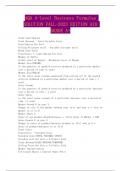
John Sulston received a Nobel prize for finding the cell lineage of the C. elegans. It shows the exact
cell divisions of the C. elegans from the first cell till it becomes an adult.
= cell
lineage of C.
elegans
Stem cell divisions
1
, C. ELEGANS INTRO
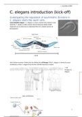
C. elegans has seam cells which work like stem cells.
The C. elegans has 4 larva stages
after it hatched from the egg. In
these stages (L1-L4) the seam
cells divide. There are 2 types of
these divisions:
asymmetrical stem cell-
like divisions
symmetrical proliferate
divisions.
The asymmetrical divisions are
shown in the image by 2 lines
who are not of the same length.
One cell has differentiated into a
hypodermal cell for example, the
other cell stays a seam cell.
The symmetrical divisions are the bold lines in the image. These lines are both of the same length,
which means that the seam cell divided into 2 new seam cells to proliferate.
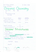
Worms hatch with 10 seam cells.
Here is a worm shown after a few asymmetrical divisions. The blue cells are new formed hypodermal
cells.
2
C. elegans introduction (kick-off)
Investigating the regulation of asymmetric divisions in
C. elegans stem-like seam cells
Caenorhabditis elegans = C. elegans is a worm used for the research in this
semester. C. elegans is often used in research because simple model
organisms are an ideal way to understand the very complex process of
development.
John Sulston received a Nobel prize for finding the cell lineage of the C. elegans. It shows the exact
cell divisions of the C. elegans from the first cell till it becomes an adult.
= cell
lineage of C.
elegans
Stem cell divisions
1
, C. ELEGANS INTRO
C. elegans has seam cells which work like stem cells.
The C. elegans has 4 larva stages
after it hatched from the egg. In
these stages (L1-L4) the seam
cells divide. There are 2 types of
these divisions:
asymmetrical stem cell-
like divisions
symmetrical proliferate
divisions.
The asymmetrical divisions are
shown in the image by 2 lines
who are not of the same length.
One cell has differentiated into a
hypodermal cell for example, the
other cell stays a seam cell.
The symmetrical divisions are the bold lines in the image. These lines are both of the same length,
which means that the seam cell divided into 2 new seam cells to proliferate.
Worms hatch with 10 seam cells.
Here is a worm shown after a few asymmetrical divisions. The blue cells are new formed hypodermal
cells.
2