PULMONARY EMBOLISM
Patho
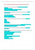
Obstruction of one or more of the branches of the pulmonary artery by particulate
matter that has an origin elsewhere in the body.
A pulmonary embolus is most caused by a thrombus. Can be a piece of tumor,
amniotic fluid, air, or fat, referred to as a no thrombotic pulmonary embolus
Epidemiology
Greatest risk factor is the presence of a deep vein thrombosis
Venous stasis
Vessel wall damage
Hypercoagulability is the major predisposing factor for the development of a DVT.
(The most common cause of DVT is prolonged immobility)
Clinical Manifestations
Sudden onset of
Dyspnea
Pleuritic chest pain
Tachypnea/cardia
Hypotension
Anxious, restless, and/or confused, impending doom
Pulmonary embolism should be suspected in long-bone surgeries with a new onset of
shortness of breath.
Diagnosis The diagnosis of PE is done through imaging studies and laboratory studies
Imaging Studies
ECG: Rule out a myocardial infarction (chest pain)
Chest x-ray: Rule out other causes of the respiratory distress
Computed tomography (CT) scan: with contrast is the most ordered test to
diagnose a PE.
Pulmonary angiography: most definitive study for the diagnosis of PE, allows for
visualization of the pulmonary vasculature, detection of any obstruction.
Laboratory Studies
A plasma D-dimer level is a very specific indicator of the possibility of the
presence of a thrombus in the body. A positive D-dimer indicates the presence of a
clot but requires further testing.
Patho
Obstruction of one or more of the branches of the pulmonary artery by particulate
matter that has an origin elsewhere in the body.
A pulmonary embolus is most caused by a thrombus. Can be a piece of tumor,
amniotic fluid, air, or fat, referred to as a no thrombotic pulmonary embolus
Epidemiology
Greatest risk factor is the presence of a deep vein thrombosis
Venous stasis
Vessel wall damage
Hypercoagulability is the major predisposing factor for the development of a DVT.
(The most common cause of DVT is prolonged immobility)
Clinical Manifestations
Sudden onset of
Dyspnea
Pleuritic chest pain
Tachypnea/cardia
Hypotension
Anxious, restless, and/or confused, impending doom
Pulmonary embolism should be suspected in long-bone surgeries with a new onset of
shortness of breath.
Diagnosis The diagnosis of PE is done through imaging studies and laboratory studies
Imaging Studies
ECG: Rule out a myocardial infarction (chest pain)
Chest x-ray: Rule out other causes of the respiratory distress
Computed tomography (CT) scan: with contrast is the most ordered test to
diagnose a PE.
Pulmonary angiography: most definitive study for the diagnosis of PE, allows for
visualization of the pulmonary vasculature, detection of any obstruction.
Laboratory Studies
A plasma D-dimer level is a very specific indicator of the possibility of the
presence of a thrombus in the body. A positive D-dimer indicates the presence of a
clot but requires further testing.