Lecture 6 20th February 2017
Neural Circuits
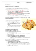
Control of motor function by cortical circuits
The corticospinal tracts: direct pathways from the motor cortex to lower
neurons (appreciate don't remember)
Neurons in the motor cortex give rise to axons that travel through the
internal capsule to the ventral surface of the midbrain.
These axons continue through the pons and come to lie on the ventral
surface of the medulla, giving rise to the pyramids.
Most of these pyramidal fibres cross in the caudal part of the medulla to
form the lateral corticospinal tract in the spinal cord.
These descending pathways play a key role in the planning, initiation,
execution and direction of voluntary movements
Topographic map of the body musculature in the primary motor cortex: a early
and rather simplistic view
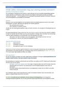
The motor cortex has been further subdivided
into functionally distinct areas
The primary motor cortex (M1), the
premotor area (PMA) and supplementary
motor area (SMA) in the human cerebral
cortex.
The primary motor cortex is located in the
precentral gyrus; the supplementary and
premotor areas are more rostral.
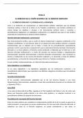
Five categories of ethologically relevant
movements evoked by ‘meaningful’ (long)
electrical stimulation in the primary motor cortex
Defensive like posture of face
Hand to mouth
Manipulation-like shaping of fingers (precision grip) and movement of
hand to central space
Outward reach with hand opened as if shaping to grasp
Climbing or leaping like posture involving all four limbs
Primary motor cortex is involved in a lot more than just making
muscles twitch
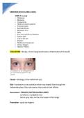
Elucidating the role of different areas of the motor cortex by recording the
activity of single neurons in awake primates
- Record the same neurons at certain tasks
Experimental apparatus developed to record the activity of (multiple)
single neurons in awake primates trained to perform specific movements
The neuron’s activity depends on a particular direction
Populations of single neurons in the motor cortex are directionally tuned
to different angles of movement
Neural Circuits
Control of motor function by cortical circuits
The corticospinal tracts: direct pathways from the motor cortex to lower
neurons (appreciate don't remember)
Neurons in the motor cortex give rise to axons that travel through the
internal capsule to the ventral surface of the midbrain.
These axons continue through the pons and come to lie on the ventral
surface of the medulla, giving rise to the pyramids.
Most of these pyramidal fibres cross in the caudal part of the medulla to
form the lateral corticospinal tract in the spinal cord.
These descending pathways play a key role in the planning, initiation,
execution and direction of voluntary movements
Topographic map of the body musculature in the primary motor cortex: a early
and rather simplistic view
The motor cortex has been further subdivided
into functionally distinct areas
The primary motor cortex (M1), the
premotor area (PMA) and supplementary
motor area (SMA) in the human cerebral
cortex.
The primary motor cortex is located in the
precentral gyrus; the supplementary and
premotor areas are more rostral.
Five categories of ethologically relevant
movements evoked by ‘meaningful’ (long)
electrical stimulation in the primary motor cortex
Defensive like posture of face
Hand to mouth
Manipulation-like shaping of fingers (precision grip) and movement of
hand to central space
Outward reach with hand opened as if shaping to grasp
Climbing or leaping like posture involving all four limbs
Primary motor cortex is involved in a lot more than just making
muscles twitch
Elucidating the role of different areas of the motor cortex by recording the
activity of single neurons in awake primates
- Record the same neurons at certain tasks
Experimental apparatus developed to record the activity of (multiple)
single neurons in awake primates trained to perform specific movements
The neuron’s activity depends on a particular direction
Populations of single neurons in the motor cortex are directionally tuned
to different angles of movement