B.M2 DEMONSTRATE SKILFUL PREPARATION OF MICROSCOPIC SLIDES TO OBSERVE AND DRAW THE STAGES OF MITOSIS AND
MEIOSIS.
PREPARATION OF STAINED SQUASHES OF CELLS FROM PLANT ROOT TIPS; SET-UP AND USE OF AN OPTICAL MICROSCOPE TO
IDENTIFY THE STAGES OF MITOSIS IN THESE STAINED SQUASHES.
ROOT TIP SQUASH USING GARLIC ROOT MERISTEM TISSUE.
This experiment was carried out to prepare a microscopic slide of the meristem tissue of a garlic root. A stain (toluidine blue) will
be added to the root tip so chromosomes can be observed under a microscope to see and identify the different stages in
meiosis. The following magnification X10, X20, and X40 we used.
EQUIPMENT:
100ml Beaker Filter paper
10 ml of Hydrochloric acid (5 mol dm-3) Mounted needle
Bench mat Distilled water
Paper towel Watch glass
Microscope and light source Root tip of garlic
Microscopic slide and cover slip Forceps
Toluidine blue stain Scalpel
METHODOLOGY:
I. Place a beaker on a bench mat and add 10ml of hydrochloric acid.
II. Measure 2cm of garlic root tip using a ruler and then cut it by using a scalpel. Cut the root tip before putting it into the acid
so that any reactions such as cell division from the cells will stop.
III. Place the 2cm root tip into the beaker of hydrochloric acid and leave it for 15 minutes.
IV. During the 15 minutes set up the microscope, and light source.
V. After the 15 minutes rinse the root tip in distilled water in the watch glass.
VI. Cut off 1mm of root tip and place it on to a microscopic slide.
VII. Soak the root tip in a small amount of toluidine blue stain and macerate with a mounted needle to separate the cells until
the tissue is well broken and the cells are stained dark blue.
VIII. Add a cover slip (to exclude air bubbles from the specimen) and gently apply pressure using a finger and spread and blot the
material at the same time by using a folded filter paper between the finger and slide.
IX. Observe the slide under the microscope and identify the cells undergoing mitosis. Chromosomes should stain dark blue.
HEALTH AND SAFETY:
HYDROCHLORIC ACID Corrosive and should be handled with caution, eye protection must be worn. Do not carry the beaker
with hydrochloric acid in it.
SCALPEL Both have sharp edges and can poke so make sure they are handled with care and not played about
MOUNTED NEEDLE with.
MICROSCOPE Make sure the microscope is away from the edge of the table, so that it doesn’t fall and break.
Damaging the eye piece and lens.
SKILLS
SKILLS WHICH I HAVE GAINED:
Being able to use an optical microscope by adjusting the lens to see the different stages of mitosis and meiosis more
clearly, and to bring about the focus, which allows observation of them and drawings to be sketched.
Being able to draw sketches of the different stages by observing them through a microscopic lens.
To observe the specimen under different magnifications (X10, X20, X40) to obtain a field of view, by adjusting and
focusing the microscope by Manoeuvring the focus piece and stage.
To be able to cut 1mm of root tip using a scalpel and mounted needle.
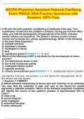
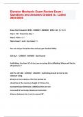
, OBSERVATIONS AND MICROSCOPIC DRAWINGS
MITOSIS:
MEIOSIS.
PREPARATION OF STAINED SQUASHES OF CELLS FROM PLANT ROOT TIPS; SET-UP AND USE OF AN OPTICAL MICROSCOPE TO
IDENTIFY THE STAGES OF MITOSIS IN THESE STAINED SQUASHES.
ROOT TIP SQUASH USING GARLIC ROOT MERISTEM TISSUE.
This experiment was carried out to prepare a microscopic slide of the meristem tissue of a garlic root. A stain (toluidine blue) will
be added to the root tip so chromosomes can be observed under a microscope to see and identify the different stages in
meiosis. The following magnification X10, X20, and X40 we used.
EQUIPMENT:
100ml Beaker Filter paper
10 ml of Hydrochloric acid (5 mol dm-3) Mounted needle
Bench mat Distilled water
Paper towel Watch glass
Microscope and light source Root tip of garlic
Microscopic slide and cover slip Forceps
Toluidine blue stain Scalpel
METHODOLOGY:
I. Place a beaker on a bench mat and add 10ml of hydrochloric acid.
II. Measure 2cm of garlic root tip using a ruler and then cut it by using a scalpel. Cut the root tip before putting it into the acid
so that any reactions such as cell division from the cells will stop.
III. Place the 2cm root tip into the beaker of hydrochloric acid and leave it for 15 minutes.
IV. During the 15 minutes set up the microscope, and light source.
V. After the 15 minutes rinse the root tip in distilled water in the watch glass.
VI. Cut off 1mm of root tip and place it on to a microscopic slide.
VII. Soak the root tip in a small amount of toluidine blue stain and macerate with a mounted needle to separate the cells until
the tissue is well broken and the cells are stained dark blue.
VIII. Add a cover slip (to exclude air bubbles from the specimen) and gently apply pressure using a finger and spread and blot the
material at the same time by using a folded filter paper between the finger and slide.
IX. Observe the slide under the microscope and identify the cells undergoing mitosis. Chromosomes should stain dark blue.
HEALTH AND SAFETY:
HYDROCHLORIC ACID Corrosive and should be handled with caution, eye protection must be worn. Do not carry the beaker
with hydrochloric acid in it.
SCALPEL Both have sharp edges and can poke so make sure they are handled with care and not played about
MOUNTED NEEDLE with.
MICROSCOPE Make sure the microscope is away from the edge of the table, so that it doesn’t fall and break.
Damaging the eye piece and lens.
SKILLS
SKILLS WHICH I HAVE GAINED:
Being able to use an optical microscope by adjusting the lens to see the different stages of mitosis and meiosis more
clearly, and to bring about the focus, which allows observation of them and drawings to be sketched.
Being able to draw sketches of the different stages by observing them through a microscopic lens.
To observe the specimen under different magnifications (X10, X20, X40) to obtain a field of view, by adjusting and
focusing the microscope by Manoeuvring the focus piece and stage.
To be able to cut 1mm of root tip using a scalpel and mounted needle.
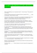
, OBSERVATIONS AND MICROSCOPIC DRAWINGS
MITOSIS: