🟥
The Gastrointestinal System
Monday, 9th May 2022
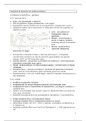
Functions of the GIT:
The GIT has 5 main functions:
1. Movement through the gut (motility) ingestion, mastication, deglutition
(swallowing) and peristalsis.
2. Secretion of enzymes and ions (as well as mucus, acid and bile) to digest
food, as well as hormones to coordinate these processes.
3. Digestion which is the catabolic breakdown of large food particles.
4. Absorption of these smaller food particles up into the blood and lymph.
5. Storage and elimination of waste products.
The main tract consists of the mouth, oesophagus, stomach, small and large
intestines and rectum.
There are also several accessory organs, like the teeth, tongue, salivary glands,
liver, gallbladder and pancreas.
The Gastrointestinal System 1
, Sphincters maintain the forward direction of digest to prevent it moving backwards or bringing
harmful secretions to the wrong areas. The GIT is around 9m in length and folds and compacts into
the abdomen. To calculate, you times the body height by 5.
The mucosa is the first layer and is the most exposed to the lumen.
Has folds and crypts (indents into the tissue layer) as well as cellular
extension called villi.
The epithelium is the highest part and secretes the mucus that protects
the GIT.
The lamina propria contains blood vessels and nerves, as well as
immune c ells.
The Gastrointestinal System 2
, The muscularis mucosae is a muscular layer that aids in peristalsis.
The submucosa is the second layer and sits below the mucosa.
The submucosa consists of lymph nodes, blood vessels and elastic collagen
fibres which allows for the stretching of the GIT but its shape maintenance.
It has (parasympathetic) nervous innervation of the Meissner’s plexus.
The muscularis layer is a muscular layer.
Two layers, the circular and longitudinal layers.
They have nerve innervation or the Auerbach’s plexus.
Responsible for the action of peristalsis.
The final level is the adventitia and is a thick layer of connective tissue that hold
the viscerae together, as well as a mucous layer to help prevent friction with
other layers.
Peyer’s patches are lymphoid follicles that run mostly in the small intestine and
facilitates the generation of the immune response against pathogens in the food
ingested.
Brunner’s glands are found in the submucosa and are responsible for the
production of mucous, and when chyme is present, mucous is secreted to
protect the duodenum to prevent against the high acidity of the stomach
contents, as well as bicarbonate ions for neutralisation.
Eurogastron prevents the secretion of HCL and.
The Crypts of Lieberkühn are boreholes that lead to intestinal glands near to the
villi.
The Oral Cavity:
There are two types of digestion: physical and chemical.
Physically, teeth aid in mastication to decrease food particles, while the tongue
assists in mastication as well as deglutination.
Chemically, glands secrete different
fluids and enzymes.
The Parotid gland (serous)
secretes 25% of the saliva
The Gastrointestinal System 3
The Gastrointestinal System
Monday, 9th May 2022
Functions of the GIT:
The GIT has 5 main functions:
1. Movement through the gut (motility) ingestion, mastication, deglutition
(swallowing) and peristalsis.
2. Secretion of enzymes and ions (as well as mucus, acid and bile) to digest
food, as well as hormones to coordinate these processes.
3. Digestion which is the catabolic breakdown of large food particles.
4. Absorption of these smaller food particles up into the blood and lymph.
5. Storage and elimination of waste products.
The main tract consists of the mouth, oesophagus, stomach, small and large
intestines and rectum.
There are also several accessory organs, like the teeth, tongue, salivary glands,
liver, gallbladder and pancreas.
The Gastrointestinal System 1
, Sphincters maintain the forward direction of digest to prevent it moving backwards or bringing
harmful secretions to the wrong areas. The GIT is around 9m in length and folds and compacts into
the abdomen. To calculate, you times the body height by 5.
The mucosa is the first layer and is the most exposed to the lumen.
Has folds and crypts (indents into the tissue layer) as well as cellular
extension called villi.
The epithelium is the highest part and secretes the mucus that protects
the GIT.
The lamina propria contains blood vessels and nerves, as well as
immune c ells.
The Gastrointestinal System 2
, The muscularis mucosae is a muscular layer that aids in peristalsis.
The submucosa is the second layer and sits below the mucosa.
The submucosa consists of lymph nodes, blood vessels and elastic collagen
fibres which allows for the stretching of the GIT but its shape maintenance.
It has (parasympathetic) nervous innervation of the Meissner’s plexus.
The muscularis layer is a muscular layer.
Two layers, the circular and longitudinal layers.
They have nerve innervation or the Auerbach’s plexus.
Responsible for the action of peristalsis.
The final level is the adventitia and is a thick layer of connective tissue that hold
the viscerae together, as well as a mucous layer to help prevent friction with
other layers.
Peyer’s patches are lymphoid follicles that run mostly in the small intestine and
facilitates the generation of the immune response against pathogens in the food
ingested.
Brunner’s glands are found in the submucosa and are responsible for the
production of mucous, and when chyme is present, mucous is secreted to
protect the duodenum to prevent against the high acidity of the stomach
contents, as well as bicarbonate ions for neutralisation.
Eurogastron prevents the secretion of HCL and.
The Crypts of Lieberkühn are boreholes that lead to intestinal glands near to the
villi.
The Oral Cavity:
There are two types of digestion: physical and chemical.
Physically, teeth aid in mastication to decrease food particles, while the tongue
assists in mastication as well as deglutination.
Chemically, glands secrete different
fluids and enzymes.
The Parotid gland (serous)
secretes 25% of the saliva
The Gastrointestinal System 3