Notas de lectura
Pharmacy Human Biology
- Grado
- Institución
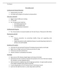
This document covers all parts of the human body in great depth. Along with signs & symptoms of certain illnesses relating to that body part and how to treat them
[Mostrar más]Vista previa 4 fuera de 227 páginas
Vista previa 4 fuera de 227 páginas


Compradores de Stuvia evaluaron más de 700.000 resúmenes. Así estas seguro que compras los mejores documentos!

Puedes pagar rápidamente y en una vez con iDeal, tarjeta de crédito o con tu crédito de Stuvia. Sin tener que hacerte miembro.

Tus compañeros escriben los resúmenes. Por eso tienes la seguridad que tienes un resumen actual y confiable. Así llegas a la conclusión rapidamente!
You get a PDF, available immediately after your purchase. The purchased document is accessible anytime, anywhere and indefinitely through your profile.
Nuestra garantía de satisfacción le asegura que siempre encontrará un documento de estudio a tu medida. Tu rellenas un formulario y nuestro equipo de atención al cliente se encarga del resto.
Stuvia is a marketplace, so you are not buying this document from us, but from seller arcane. Stuvia facilitates payment to the seller.
No, you only buy this summary for 10,26 €. You're not tied to anything after your purchase.
4.6 stars on Google & Trustpilot (+1000 reviews)
45,681 summaries were sold in the last 30 days
Founded in 2010, the go-to place to buy summaries for 14 years now