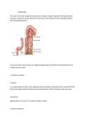
THE RECTUM
The rectum is the final, straight (from Latin rectus, meaning "straight") segment of the large intestine,
serving as a temporary storage chamber for feces and a crucial conduit for their controlled expulsion
from the body (defecation).
It is far more than a passive tube; it is a highly specialized organ with distinct anatomical features and
complex nervous control.
1. Anatomy & Location
· Position:
It is located within the pelvic cavity, beginning at the termination of the sigmoid colon at the level of the
third sacral vertebra (S3) and ending at the anorectal junction, where it continues as the anal canal.
· Dimensions:
Approximately 12-15 cm (4.7-5.9 inches) in length in adults.
· Anatomical Features:
, · Rectal Ampulla:
The lower, dilated portion of the rectum. Its walls are highly distensible, allowing it to act as a reservoir
for fecal material until defecation is socially convenient.
This is its primary storage function.
· Rectal Valves (of Houston): These are 2-4 permanent, transverse folds of the rectal mucosa (inner
lining).
They are not valves in the traditional sense but serve important functions:
1. They support the weight of fecal matter.
2. They slow the passage of feces, allowing for further absorption of water and electrolytes.
3. They separate flatus (gas) from solid stool, allowing gas to pass without the simultaneous passage
of feces.
· Peritoneal Relations:
The upper third of the rectum is covered by peritoneum on its front and sides, the middle third only on
the front, and the lower third is completely extraperitoneal (behind the peritoneum).
This is surgically important.
· Muscular Layers:
Like the rest of the GI tract, it has an inner circular muscle layer and an outer longitudinal layer.
These layers are thickened at the junction with the anal canal to form the internal anal sphincter
(involuntary, smooth muscle).
2. Histology (Microscopic Anatomy)
The rectal wall consists of the standard four layers of the GI tract: