

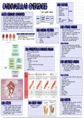
CARDIOVASCLILAR EMERGENCIES RISK FACTORS
>
-
>
-
>
-
obesity
lask of physical activity
hypertension -
modifiable
ACUTE CARANARY SYNDROME >
-
smoking
ACUTE CORONARY SYNDROMES >
hyperlipidemia
-
>
-
diabetes mellitus
-
acute myocardial infarction (AMI) Sudden cardias death /SCD) sire amang the
most serious catastrophic of acute cardias disorders accounting for
hundreds of thousands of deaths each year worldwide
, ANGINA YNSTABLE NSTEMI STEM/ >
-
aging
non-
> genetic predisposition
-
-
modifiable
-
in most cases of AMI 3 in a majority of cases of SCD the underlying >
-
male gender
,
>
pathology is acute intraluminal coronary thrombus formation within a coronary LOW RISK HIGH RISK
artery leading to tatal or near-total acute caponary occlusion
,
CAD -STALEANGIMaes
-
the underlying pathophysiologis mechanisms for these syndromes begin with the
process of atherosclerosis which develops progresses for years prico
,
to the
a sure event
CAD-ANGINA PECTORIS from
part of the ACS spectrum
not
-
angina pectaris is most commonly caused by myocardial
-
symptoms usually occur at a predictable level of
WHAT IS CORONARY ARTERY DISEASE (CAD) ? isshaemia which results from a mismatch between exertion : O demand) O supply to the
-
narrowing/blackage of the
saronary arteries myocardial bload flow (supply) Oz demand myocardium
caused by atherosclerosis -
Cam be divided into 3 categories : -
caused by a fixed calcifiedatherosclerotic
-
can lead to
Angina or Myacardial Infarction >
-
Stable plaque that does not completely occlude the
> unstable coronary vessel I still allow blood flow
-
>
-
prinzmetal
SIGNS 3 SYMPTOMS
P : occurs with stress/exection ; relieved byoest/nitrates
substernal crushing or burning Smight think indigestion)
i
:
,
CAD-PRINIMETAL'S (VARIANT) ANGINA may radiate to shoulder back jaw
:
,
: mild to moderate pain
caused lasts = 2-5 minutes
by coronary artery
:
spasm
-
-
mild tachycardia
very rars
-
often while anxious
at night sleeping
-
-
occurs
classical normal
pts are presaminantly younger women who
may not have the ECG
-
-
.
cardiovascular risk factors
-
caused by :
>
-
>
-
exposure
stress
to said weather
CAD LINSTABLE ANGINA -
>
-
meds. that tighten/narrow blood vessels
-
forms part of the ACS spectrum
>
-
smaking
-
caused by a ruptured
plaque formingI thoombus ,
>
-
cacaine use
>
-
this causes a partial occlusion of the coronary
relieved dotery leading toa blood flow 3 Ischaemia
-
by nitoates ,
>
-
no necrosis/infarct
SIGNS i SYMPTOMS
p. at rest , sleeping or minimal exertion ; not relieved by rest
or nitoates
substernal , crushing
: may
· radiate to shoulder ,
back jaw
anis
. moderatetosquee
5
-
tachycardia
-
anxious
dyspnea
-
-
cold isalammy
-
nauseg 3 vomiting
·
ESG Changes
CA
CAD-STEMI
NO
are
very similar ,
with NSTEMI
biomar as
-
caused by a ruptured plaque farming
, a thrombus -
caused by a
ruptured plaque , forming a thrombus
>
-
causes complete osclusion of the coronary
a >
-
causes a partial
osclusion of the coronary
artery,
artary leading to no blood flow ischaemia
, leading to↓ blood flaw ischaemia
> nesoosis/ infarst
-
of transmural myosardium >
-
necrosis/infarst of subendocardial myocardium
-
ECG findings include ST elevation , new pathological
-
ECG findings often include ST depression but do not
& waves abnormal T waves include new pathological a waves
, THE 12-LEAD ECG ECGGGESCTM
-
ST
>
-
septal (V1-2)
> anterior SV3-4)
-
in contiguous lea.
> lateral
-
(13aVL , US-6/
>
-
inferior (II .I aVF)
,
>
-
right ventricular /VI ,
V4R)
>
-
posterior (VT-a)
V
ABNORMAL T WAVES [
WHAT DO WE MEAN WHEN WE SAY LEADS ARE CONTIGLOUS ?
-
contiguous leads are next to one another anatomically speaking
>
-
T-wave inversions due to myocardial ischaemia >
they view the same general area of the heart
-
ar infarction occur in contiguous leads based in addition any 2 precordial leads next to a n e another are contiguous
-
,
an the anatomical location of the area of -
in other words V43 US are sontiguous even though V4 is an anterior
. ,
is shaemia/infaction : lead & US is a lateral lead
·
inferior > this
-
makes sense when you consider that leads VLs VS are next
·
lateral to each other on the patient's chest
·
septal
· anterior
-
hyperacute
·
PATHOLOGCQ ue to acute mi
-
inferiora Waves (I ,I alf),
with T-wave inversion due to
previous Mr
CORONARY ARTERY CULPRIT
>
-
RIGHT CORONARY ARTERY
-
supplies blood to :
·
right atoium
STELLATION
·
portions of both ventrisles
·
·
SA AV mode
right ventricle
·
st elevation
-
inferior M1 with different morphology
-
right ventricle ML
-
posteriar MI
>
-
LEFT CORONARY ARTERY STEMI PROGRESSION
-
supplies bload to :
·
left ventrisle
·
left atrium
·
intraventricular septum
-
2 main branches :
·
left anterior descending
Sircumflex
·
-
anterior M1 SLAD)
-
Septal (LAD)
-
lateral (airsumflex]
-
posterior 1 circumflex)
>
-
>
-
>
-
obesity
lask of physical activity
hypertension -
modifiable
ACUTE CARANARY SYNDROME >
-
smoking
ACUTE CORONARY SYNDROMES >
hyperlipidemia
-
>
-
diabetes mellitus
-
acute myocardial infarction (AMI) Sudden cardias death /SCD) sire amang the
most serious catastrophic of acute cardias disorders accounting for
hundreds of thousands of deaths each year worldwide
, ANGINA YNSTABLE NSTEMI STEM/ >
-
aging
non-
> genetic predisposition
-
-
modifiable
-
in most cases of AMI 3 in a majority of cases of SCD the underlying >
-
male gender
,
>
pathology is acute intraluminal coronary thrombus formation within a coronary LOW RISK HIGH RISK
artery leading to tatal or near-total acute caponary occlusion
,
CAD -STALEANGIMaes
-
the underlying pathophysiologis mechanisms for these syndromes begin with the
process of atherosclerosis which develops progresses for years prico
,
to the
a sure event
CAD-ANGINA PECTORIS from
part of the ACS spectrum
not
-
angina pectaris is most commonly caused by myocardial
-
symptoms usually occur at a predictable level of
WHAT IS CORONARY ARTERY DISEASE (CAD) ? isshaemia which results from a mismatch between exertion : O demand) O supply to the
-
narrowing/blackage of the
saronary arteries myocardial bload flow (supply) Oz demand myocardium
caused by atherosclerosis -
Cam be divided into 3 categories : -
caused by a fixed calcifiedatherosclerotic
-
can lead to
Angina or Myacardial Infarction >
-
Stable plaque that does not completely occlude the
> unstable coronary vessel I still allow blood flow
-
>
-
prinzmetal
SIGNS 3 SYMPTOMS
P : occurs with stress/exection ; relieved byoest/nitrates
substernal crushing or burning Smight think indigestion)
i
:
,
CAD-PRINIMETAL'S (VARIANT) ANGINA may radiate to shoulder back jaw
:
,
: mild to moderate pain
caused lasts = 2-5 minutes
by coronary artery
:
spasm
-
-
mild tachycardia
very rars
-
often while anxious
at night sleeping
-
-
occurs
classical normal
pts are presaminantly younger women who
may not have the ECG
-
-
.
cardiovascular risk factors
-
caused by :
>
-
>
-
exposure
stress
to said weather
CAD LINSTABLE ANGINA -
>
-
meds. that tighten/narrow blood vessels
-
forms part of the ACS spectrum
>
-
smaking
-
caused by a ruptured
plaque formingI thoombus ,
>
-
cacaine use
>
-
this causes a partial occlusion of the coronary
relieved dotery leading toa blood flow 3 Ischaemia
-
by nitoates ,
>
-
no necrosis/infarct
SIGNS i SYMPTOMS
p. at rest , sleeping or minimal exertion ; not relieved by rest
or nitoates
substernal , crushing
: may
· radiate to shoulder ,
back jaw
anis
. moderatetosquee
5
-
tachycardia
-
anxious
dyspnea
-
-
cold isalammy
-
nauseg 3 vomiting
·
ESG Changes
CA
CAD-STEMI
NO
are
very similar ,
with NSTEMI
biomar as
-
caused by a ruptured plaque farming
, a thrombus -
caused by a
ruptured plaque , forming a thrombus
>
-
causes complete osclusion of the coronary
a >
-
causes a partial
osclusion of the coronary
artery,
artary leading to no blood flow ischaemia
, leading to↓ blood flaw ischaemia
> nesoosis/ infarst
-
of transmural myosardium >
-
necrosis/infarst of subendocardial myocardium
-
ECG findings include ST elevation , new pathological
-
ECG findings often include ST depression but do not
& waves abnormal T waves include new pathological a waves
, THE 12-LEAD ECG ECGGGESCTM
-
ST
>
-
septal (V1-2)
> anterior SV3-4)
-
in contiguous lea.
> lateral
-
(13aVL , US-6/
>
-
inferior (II .I aVF)
,
>
-
right ventricular /VI ,
V4R)
>
-
posterior (VT-a)
V
ABNORMAL T WAVES [
WHAT DO WE MEAN WHEN WE SAY LEADS ARE CONTIGLOUS ?
-
contiguous leads are next to one another anatomically speaking
>
-
T-wave inversions due to myocardial ischaemia >
they view the same general area of the heart
-
ar infarction occur in contiguous leads based in addition any 2 precordial leads next to a n e another are contiguous
-
,
an the anatomical location of the area of -
in other words V43 US are sontiguous even though V4 is an anterior
. ,
is shaemia/infaction : lead & US is a lateral lead
·
inferior > this
-
makes sense when you consider that leads VLs VS are next
·
lateral to each other on the patient's chest
·
septal
· anterior
-
hyperacute
·
PATHOLOGCQ ue to acute mi
-
inferiora Waves (I ,I alf),
with T-wave inversion due to
previous Mr
CORONARY ARTERY CULPRIT
>
-
RIGHT CORONARY ARTERY
-
supplies blood to :
·
right atoium
STELLATION
·
portions of both ventrisles
·
·
SA AV mode
right ventricle
·
st elevation
-
inferior M1 with different morphology
-
right ventricle ML
-
posteriar MI
>
-
LEFT CORONARY ARTERY STEMI PROGRESSION
-
supplies bload to :
·
left ventrisle
·
left atrium
·
intraventricular septum
-
2 main branches :
·
left anterior descending
Sircumflex
·
-
anterior M1 SLAD)
-
Septal (LAD)
-
lateral (airsumflex]
-
posterior 1 circumflex)


