
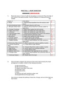
PRACTICAL 1 : HEART DISSECTION
WORKSHEET (MEMORANDUM)
1) Match the terms in Column A with the descriptions in Column B. Place the letter of
your choice in the space provided. Use your textbook for a reference source if
needed. (20)
Column A COLUMN B B
A. Aorta 1. Prevents blood backflow from left ventricle to left 4
atrium
B. Aortic Semilunar Valve 2. Transports blood to LEFT lung 7
C. Bicuspid (mitral) Valve 3. Returns venous blood from coronary system to 1
right atrium
D. Chordae Tendineae 4. Major artery leading from left ventricle 13
E. Inferior Vena Cava 5. Major vein leading into lower right atrium 5
F. Left Atrium 6. Major vein leading into upper right atrium 10
G. Left Pulmonary Artery 7. Prevents blood backflow from aorta to left ventricle 2
H. Left Ventricle 8. Pumps blood to pulmonary artery 8
I. Myocardium 9. Major artery leading from right ventricle 20
J. Coronary Arteries 10. Pumps blood to left ventricle 15
K. Coronary Sinus 11. Major veins leading into left atrium 3
L. Papillary Muscles 12. Prevents blood backflow from right ventricle to 16
right atrium
M. Pulmonary Artery (trunk) 13. Attach cuspid valve cusps to papillary muscles 9
N. Pulmonic Semilunar 14. Pumps blood to right ventricle 19
Valve
O. Pulmonary Veins 15. Transport blood from aorta to cardiac muscles 11
P. Right Atrium 16. Anchor sites for the chordae tendineae 14
Q. Right Ventricle 17. Pumps blood to aorta 17
R. Superior Vena Cava 18. Myocardium that separates the two ventricles 6
S. Tricuspid Valve 19. Prevents blood backflow from pulmonary artery to 12
right ventricle.
T. Ventricular Septum 20. Muscle wall of the heart 18
2) Draw and label a diagram the structure of the human heart showing the proper
arrangement or location of EACH of the following structures:
A) right and left atria;
B) right and left ventricles;
C) superior and inferior vena cava;
D) pulmonary veins;
E) pulmonary artery;
F) aorta;
G) pulmonic and aortic semilunar valves; and,
H) bicuspid/mitral and tricuspid valves.
Use arrows to indicate the pathway of blood into, through, and out of the heart you
have drawn. (17)
WORKSHEET (MEMORANDUM)
1) Match the terms in Column A with the descriptions in Column B. Place the letter of
your choice in the space provided. Use your textbook for a reference source if
needed. (20)
Column A COLUMN B B
A. Aorta 1. Prevents blood backflow from left ventricle to left 4
atrium
B. Aortic Semilunar Valve 2. Transports blood to LEFT lung 7
C. Bicuspid (mitral) Valve 3. Returns venous blood from coronary system to 1
right atrium
D. Chordae Tendineae 4. Major artery leading from left ventricle 13
E. Inferior Vena Cava 5. Major vein leading into lower right atrium 5
F. Left Atrium 6. Major vein leading into upper right atrium 10
G. Left Pulmonary Artery 7. Prevents blood backflow from aorta to left ventricle 2
H. Left Ventricle 8. Pumps blood to pulmonary artery 8
I. Myocardium 9. Major artery leading from right ventricle 20
J. Coronary Arteries 10. Pumps blood to left ventricle 15
K. Coronary Sinus 11. Major veins leading into left atrium 3
L. Papillary Muscles 12. Prevents blood backflow from right ventricle to 16
right atrium
M. Pulmonary Artery (trunk) 13. Attach cuspid valve cusps to papillary muscles 9
N. Pulmonic Semilunar 14. Pumps blood to right ventricle 19
Valve
O. Pulmonary Veins 15. Transport blood from aorta to cardiac muscles 11
P. Right Atrium 16. Anchor sites for the chordae tendineae 14
Q. Right Ventricle 17. Pumps blood to aorta 17
R. Superior Vena Cava 18. Myocardium that separates the two ventricles 6
S. Tricuspid Valve 19. Prevents blood backflow from pulmonary artery to 12
right ventricle.
T. Ventricular Septum 20. Muscle wall of the heart 18
2) Draw and label a diagram the structure of the human heart showing the proper
arrangement or location of EACH of the following structures:
A) right and left atria;
B) right and left ventricles;
C) superior and inferior vena cava;
D) pulmonary veins;
E) pulmonary artery;
F) aorta;
G) pulmonic and aortic semilunar valves; and,
H) bicuspid/mitral and tricuspid valves.
Use arrows to indicate the pathway of blood into, through, and out of the heart you
have drawn. (17)


