DNA: THE CODE OF LIFE
HISTORY OF STRUCTURE ROLE OF DNA
- Rosalind Franklin and Maurice Wilkins: 1. Carry hereditary info in each cell in the form of
X-ray crystallography. genes.
- Francis Crick and James Watson: 2. Provide blueprint for organism’s growth and
Analysed x-ray data from Rosalind and others; development by coding for protein synthesis.
built a model out of brass plates and clamps. 3. Can replicate, allow daughter cells to get a
copy of genetic info as it is passed on from
HISTORY OF GENETIC REPLICATION generation to generation.
- James Watson and Francis Crick:
Pairing of base pairs suggested that given a NON-CODING DNA
sequence of bases in one strand, other strand
automatically determined. (Each strand can - Less than 2% is coding DNA.
replicate – template). - Protein-coding regions called exons and non-
- Francis Crick and Sydney Brenner: coding regions called introns.
(Protein synthesis) Triplet code used in reading - Complex organisms have more introns.
genetic material in DNA and transferring this - Non-coding DNA form functional RNA
information from nucleus to cytoplasm via RNA, molecules which have regulatory functions.
to where proteins are made. Form is function –
replicate and carry instructions. REPLICATION
- Defined as the process of making a new DNA
NOBEL PRIZE molecule from an existing DNA molecule that is
- Rosalind only one with degrees in chemistry. identical to the original DNA molecule.
Suspected DNA molecules were helical in - Takes place during interphase.
structure, wanted further proof. Died at 37 due - Necessary to ensure that the genetic code is
to cancer. passed on to each new daughter cell formed
- 1962; Watson, Crick and Wilkins won Nobel Prize during cell division.
for physiology/medicine. 1. Process catalysed by enzyme DNA polymerase.
2. Double helix unwinds.
WHERE IS DNA FOUND? 3. Weak hydrogen bonds break between bases,
allowing strand to part.
- Mainly in nucleus; forms part of chromosomes 4. Each single chain of DNA is exposed.
that make up chromatin network. 5. Free nucleotides become attached to their
- Chromatin is chromosomal material made up of matching, exposed base partners.
DNA, RNA and histone proteins as found in a 6. 2 daughter DNA molecules (identical to original
non-dividing cell. due to base pairs) twist to form a double helix,
- Extranuclear/Extracellular DNA: mitochondria in which then winds itself around the histones,
plants and animals/chloroplasts in plants. forming a chromosome. Whole process takes a
couple of seconds.
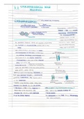
STRUCTURE OF DNA
- 2 strands twist to form a stable, 3-dimensional
double helix.
- Molecule is a long chain (polymer) made up of
small units (monomers) called nucleotides.
- Each nucleotide has a sugar molecule
(deoxyribose), a phosphate molecule and a
nitrogenous base.
o Adenine (A) + Thymine (T).
o Guanine (G) + Cytosine (C).
- Sugar joins with base.
- Between bases ➔ weak hydrogen bond.
- Between nucleotides ➔ strong covalent bond.
- 4 bases are foundation of genetic code;
instructs cells on how to synthesize enzymes and
other proteins.
- Organisms differ due to the sequence of
nucleotides that are strung together. (Sequence
of nucleotides therefore determines the genetic
code of an organism).
, RNA
STRUCTURE MITOCHONDRIAL EVE
- Polymer made up of nucleotides, differs from First point on mtDNA network where everyone would
DNA: meet.
1. Single strand.
2. Shorter strand. MTDNA TESTING
3. Sugar is ribose.
1. Scrape of tissue from inside cheek sent to any
4. Uracil instead of thymine.
lab of genetic genealogy for mtDNA testing.
2. Genetic code studied at specific locations and
FUNCTION results are compared to the sequence of
Carries instructions from DNA in nucleus to ribosomes in mtDNA of other individuals or of the reference
cytoplasm of a cell where it controls the synthesis of sample available in the lab. (CRS – Cambridge
proteins from amino acids. Reference Sequence; source of info of human
mtDNA commonly found in people of European
Similarities between DNA and RNA descent).
1. Both made of up of polymers, nucleotides 3. Similar genetic sequences in mtDNA testing
consisting of a sugar, phosphate and base, 4 indicate a common female ancestor.
bases.
2. Both responsible for synthesis of proteins. HOW CAN MTDNA PROFILES BE USED?
1. Reconstruct family maternal-linked relationships.
MITOCHONDRIAL DNA STRUCTURE 2. Investigate forensic cases where chromosomal
- Double-stranded, ring-shaped molecule found DNA is degraded.
in all mitochondria. 3. Determine if siblings have the same mother.
- Entirely inherited from mother (comes from
oocyte).
- Has its own genome of about 16 500 base pairs
that code for proteins (enzymes), tRNA and
rRNA. Therefore much shorter than
chromosomal DNA. The genes are essential for
the normal functioning of mitochondria, as they
code for the enzymes that control cellular
respiration.
WHY IS DNA IN MITOCHONDRIA?
Mitochondria were once separate organisms
(prokaryotes). At some point, entered symbiotic
relationship with eukaryotic cells through endosmosis. As
a result, mitochondria contain their own circular DNA
called mtDNA.
TRACING GENETIC LINKS
- During fertilisation, only sperm enters ovum;
therefore, all organelles come from mother.
- As mtDNA passed on from gen to gen, direct
maternal genetic line created.
- mtDNA mutates occasionally ➔ substitution can
take place where one nucleotide is replaced by
another, resulting in a site known as a marker.
- mtDNA profiles of 2 sets of markers can be
made:
o Profiles similar; organisms closely
related.
o Profiles different; organisms have
diverged along different evolutionary
pathways.
- Markers can be mapped through generations
➔ enables researchers to trace lineage through
females (matrilineage).
HISTORY OF STRUCTURE ROLE OF DNA
- Rosalind Franklin and Maurice Wilkins: 1. Carry hereditary info in each cell in the form of
X-ray crystallography. genes.
- Francis Crick and James Watson: 2. Provide blueprint for organism’s growth and
Analysed x-ray data from Rosalind and others; development by coding for protein synthesis.
built a model out of brass plates and clamps. 3. Can replicate, allow daughter cells to get a
copy of genetic info as it is passed on from
HISTORY OF GENETIC REPLICATION generation to generation.
- James Watson and Francis Crick:
Pairing of base pairs suggested that given a NON-CODING DNA
sequence of bases in one strand, other strand
automatically determined. (Each strand can - Less than 2% is coding DNA.
replicate – template). - Protein-coding regions called exons and non-
- Francis Crick and Sydney Brenner: coding regions called introns.
(Protein synthesis) Triplet code used in reading - Complex organisms have more introns.
genetic material in DNA and transferring this - Non-coding DNA form functional RNA
information from nucleus to cytoplasm via RNA, molecules which have regulatory functions.
to where proteins are made. Form is function –
replicate and carry instructions. REPLICATION
- Defined as the process of making a new DNA
NOBEL PRIZE molecule from an existing DNA molecule that is
- Rosalind only one with degrees in chemistry. identical to the original DNA molecule.
Suspected DNA molecules were helical in - Takes place during interphase.
structure, wanted further proof. Died at 37 due - Necessary to ensure that the genetic code is
to cancer. passed on to each new daughter cell formed
- 1962; Watson, Crick and Wilkins won Nobel Prize during cell division.
for physiology/medicine. 1. Process catalysed by enzyme DNA polymerase.
2. Double helix unwinds.
WHERE IS DNA FOUND? 3. Weak hydrogen bonds break between bases,
allowing strand to part.
- Mainly in nucleus; forms part of chromosomes 4. Each single chain of DNA is exposed.
that make up chromatin network. 5. Free nucleotides become attached to their
- Chromatin is chromosomal material made up of matching, exposed base partners.
DNA, RNA and histone proteins as found in a 6. 2 daughter DNA molecules (identical to original
non-dividing cell. due to base pairs) twist to form a double helix,
- Extranuclear/Extracellular DNA: mitochondria in which then winds itself around the histones,
plants and animals/chloroplasts in plants. forming a chromosome. Whole process takes a
couple of seconds.
STRUCTURE OF DNA
- 2 strands twist to form a stable, 3-dimensional
double helix.
- Molecule is a long chain (polymer) made up of
small units (monomers) called nucleotides.
- Each nucleotide has a sugar molecule
(deoxyribose), a phosphate molecule and a
nitrogenous base.
o Adenine (A) + Thymine (T).
o Guanine (G) + Cytosine (C).
- Sugar joins with base.
- Between bases ➔ weak hydrogen bond.
- Between nucleotides ➔ strong covalent bond.
- 4 bases are foundation of genetic code;
instructs cells on how to synthesize enzymes and
other proteins.
- Organisms differ due to the sequence of
nucleotides that are strung together. (Sequence
of nucleotides therefore determines the genetic
code of an organism).
, RNA
STRUCTURE MITOCHONDRIAL EVE
- Polymer made up of nucleotides, differs from First point on mtDNA network where everyone would
DNA: meet.
1. Single strand.
2. Shorter strand. MTDNA TESTING
3. Sugar is ribose.
1. Scrape of tissue from inside cheek sent to any
4. Uracil instead of thymine.
lab of genetic genealogy for mtDNA testing.
2. Genetic code studied at specific locations and
FUNCTION results are compared to the sequence of
Carries instructions from DNA in nucleus to ribosomes in mtDNA of other individuals or of the reference
cytoplasm of a cell where it controls the synthesis of sample available in the lab. (CRS – Cambridge
proteins from amino acids. Reference Sequence; source of info of human
mtDNA commonly found in people of European
Similarities between DNA and RNA descent).
1. Both made of up of polymers, nucleotides 3. Similar genetic sequences in mtDNA testing
consisting of a sugar, phosphate and base, 4 indicate a common female ancestor.
bases.
2. Both responsible for synthesis of proteins. HOW CAN MTDNA PROFILES BE USED?
1. Reconstruct family maternal-linked relationships.
MITOCHONDRIAL DNA STRUCTURE 2. Investigate forensic cases where chromosomal
- Double-stranded, ring-shaped molecule found DNA is degraded.
in all mitochondria. 3. Determine if siblings have the same mother.
- Entirely inherited from mother (comes from
oocyte).
- Has its own genome of about 16 500 base pairs
that code for proteins (enzymes), tRNA and
rRNA. Therefore much shorter than
chromosomal DNA. The genes are essential for
the normal functioning of mitochondria, as they
code for the enzymes that control cellular
respiration.
WHY IS DNA IN MITOCHONDRIA?
Mitochondria were once separate organisms
(prokaryotes). At some point, entered symbiotic
relationship with eukaryotic cells through endosmosis. As
a result, mitochondria contain their own circular DNA
called mtDNA.
TRACING GENETIC LINKS
- During fertilisation, only sperm enters ovum;
therefore, all organelles come from mother.
- As mtDNA passed on from gen to gen, direct
maternal genetic line created.
- mtDNA mutates occasionally ➔ substitution can
take place where one nucleotide is replaced by
another, resulting in a site known as a marker.
- mtDNA profiles of 2 sets of markers can be
made:
o Profiles similar; organisms closely
related.
o Profiles different; organisms have
diverged along different evolutionary
pathways.
- Markers can be mapped through generations
➔ enables researchers to trace lineage through
females (matrilineage).