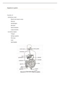
Digestive system
Consists of:
- alimentary canal
Mouth and mouth cavity
Pharynx
Oesophagus
Stomach
Small intestine
Large intestine
- accessory organs
Tongue
Salivary glands
Pancreas
Liver
Gall bladder
,structure and functions of the alimentary canal
mouth and mouth cavity
- upper opening and surrounded by two fleshy lips
- mouth cavity = inside cavity and is lined with a mucous membrane. Roof consists of a
hard ridged palate at the front and soft palate (ending in uvula – closes nasal
cavity when swallowing) at the back. Also contains the tongue and teeth
toward the front and openings of three pairs of salivary glands
functions
- receives food and begins the process of mechanical digestion by breaking down larger
food particles into smaller ones and mixing it with saliva
pharynx (throat cavity)
- joins at back of mouth cavity
- leads to two openings
oesophagus
trachea
functions
- muscles are responsible for peristalsis which pushes the bolus (round ball of chewed
food that is mixed with saliva in mouth cavity) forward
stomach
- sickle-shaped, sac-like organ located below the diaphragm
- stomach wall is thick and muscular
- at top: cardiac sphincter closes the opening to the oesophagus
- at lower: pyloric sphincter closes the opening to the small intestine
- blood transports the hormone gastrin from the stomach mucosa to the gastric glands
when food reaches the stomach to stimulate them to secret gastric juice
- gastric glands can produce excessive amounts of gastric juice under stressful
conditions, destroying the mucous lining, leading to stomach ulcers
- acidic, fluid mass can push up into the oesophagus, resulting in heartburn
functions
- muscular wall causes churning movements that assist with physical digestion and
ensures food is mixed with gastric juices
- glands secrete gastric juices
food leaves stomach in a semi-solid state called chyme
, Small intestine
- long, muscular tube approx 5-6m
- consists of three parts
Duodenum: first and shortest. Common bile duct and pancreatic duct opens as
joint tube in duodenum
Jejunum: middle part of small intestine
Ileum: last and longest. Joins to caecum (first part of large intestine), closed by a
ring of muscle (ileo-caecal sphincter)
- wall consists of four layers
Serous membrane: outer connective tissue layer
Muscle layer: outer (longitudinal muscles) and inner (circular muscles)
Submucosa: layer of connective tissue with blood vessels, lymph vessels, nerves
and glands
Mucosa: innermost layer with transverse folds containing millions of finger-like
projections on these folds
functions
- muscles in wall cause peristalsis, moving chyme forward and ensuring it mixes
thoroughly with digestive juices
- glands in duodenal wall (crypts of Lieberkühn and Brunner gland) secrete digestive juice
(intestinal juice) which play a role in digestion
- millions of villi increase the surface area for absorption of digested nutrients