Page 1
Chromosomes and Gene Maps
✽ Basic General Definitions:
- Mitosis – replicated chromosomes auto-orientate at metaphase and equationally divide at
telophase to produce two identical daughter cells
- Meiosis – replicated chromosomes co-orientate at metaphase I and reductionally divide at
telophase I; then, single chromosomes auto-orientate at metaphase II, and equationally
divide at telophase II to produce four gametes
- Gene – segments of DNA that code for a particular protein
- Chromosome – unit of tightly packed DNA
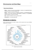
Introduction to Genetics:
Human chromosomes show the location of genes, and their abnormal forms cause hereditary
diseases (due to mutations)
diagnostic tests can find the genetic
mutations on specific chromosomes
, Page 2
Types of Chromosomes:
Chromosome Banding:
- Technique for differential or selective staining of mitotic and meiotic chromosomes to produce a
characteristic banding pattern of certain chromosomal regions
- e.g. centromeres, nucleolus organising region (NOR), and G-C or A-T rich regions
➢ The staining process occurs during metaphase as the chromosomes are highly condensed
and are thus easier to visualise
• Differential staining:
- Fluorescent and Giesma staining techniques
- Induces light and dark bands across the entire length of the chromosomes
- e.g. Quinacrine (Q), Reverse (R), and Giemsa (G) bands
• Selective staining:
- Different regions of DNA are subject to a considerable degree of stain variation
- e.g. C bands, NOR, G-11, and Cd bands (shows the active centromere)
, Page 3
Technique Procedure Banding Pattern
- Dark bands (A-T rich)
- Proteolysis with Trypsin - Light bands (G-C rich)
G- Banding
- Stained with Giemsa Dye ↳ gives light and dark stains along
the length of the chromosome
- Dark bands (G-C rich)
- Heat denatured
R- Banding - Light bands (A-T rich)
- Stained with Giemsa Dye
↳ stains non-centromeric regions
- Dark bands (A-T rich)
- Light bands (G-C rich)
Q- Banding - Stained with Quinacrine Dye
↳ yields a similar pattern to
G-banding
- Denatured with Barium - Dark bands (constitutive
C- Banding - Hydroxide heterochromatin)
- Stained with Giemsa Dye ↳ only stains the centromeres
➢ These banding techniques define the karyotype of an organism, and are used to:
- Distinguish chromosomes and regions of chromosomes
- Identify various abnormalities
- Map genes accurately
- Compare banding of related species
- Identify parental origin of different chromosomes
Karyotype vs. Idiogram:
- Karyotype = chromosome complement of a
cell or individual
- Pro-metaphase chromosomes arranged in a
sequence according to length and centro-
mere position
➢ On the karyotype, D and G groups are acrocentric chromosomes, and they all have a secondary
constriction near the end, called 'satellite DNA'
➢ The centromere is the primary constriction – it associates with the nucleolus organising region
(NOR) in the nucleus
➢ The secondary constriction converts rDNA to rRNA, as it associates with the ribosomes to
allow for protein synthesis to occur
, Page 4
- Idiogram = diagrammatic representation of the karyotype
- Pictorial reference point useful for locating positions of individual genes on chromosomes and
identifying abnormalities associated with chromosomal disorders
- Enables members of scientific community to reference important sources (e.g. Human
Genome project) through a universal vocabulary that allows for quick clear interpretation –
i.e. International System for Cytogenetic Nomenclature (ISCN)
International System for Cytogenetic Nomenclature (ISCN):
- Numbering begins at the centromere
- Chromosomes are assigned long arm and short arm
- Each arm is divided into regions
- Regions are identified by specific morphological features
- i.e. presence of Giemsa-staining bands
- Regions are subdivided into subregions
- Subregions are divided into regions
- e.g. 12q24.3
↳ 12 = chromosome 12
↳ q = long arm
↳ 2 = second region
↳ 4 = fourth band
↳ 3 = subregion 3
Chromosomes and Gene Maps
✽ Basic General Definitions:
- Mitosis – replicated chromosomes auto-orientate at metaphase and equationally divide at
telophase to produce two identical daughter cells
- Meiosis – replicated chromosomes co-orientate at metaphase I and reductionally divide at
telophase I; then, single chromosomes auto-orientate at metaphase II, and equationally
divide at telophase II to produce four gametes
- Gene – segments of DNA that code for a particular protein
- Chromosome – unit of tightly packed DNA
Introduction to Genetics:
Human chromosomes show the location of genes, and their abnormal forms cause hereditary
diseases (due to mutations)
diagnostic tests can find the genetic
mutations on specific chromosomes
, Page 2
Types of Chromosomes:
Chromosome Banding:
- Technique for differential or selective staining of mitotic and meiotic chromosomes to produce a
characteristic banding pattern of certain chromosomal regions
- e.g. centromeres, nucleolus organising region (NOR), and G-C or A-T rich regions
➢ The staining process occurs during metaphase as the chromosomes are highly condensed
and are thus easier to visualise
• Differential staining:
- Fluorescent and Giesma staining techniques
- Induces light and dark bands across the entire length of the chromosomes
- e.g. Quinacrine (Q), Reverse (R), and Giemsa (G) bands
• Selective staining:
- Different regions of DNA are subject to a considerable degree of stain variation
- e.g. C bands, NOR, G-11, and Cd bands (shows the active centromere)
, Page 3
Technique Procedure Banding Pattern
- Dark bands (A-T rich)
- Proteolysis with Trypsin - Light bands (G-C rich)
G- Banding
- Stained with Giemsa Dye ↳ gives light and dark stains along
the length of the chromosome
- Dark bands (G-C rich)
- Heat denatured
R- Banding - Light bands (A-T rich)
- Stained with Giemsa Dye
↳ stains non-centromeric regions
- Dark bands (A-T rich)
- Light bands (G-C rich)
Q- Banding - Stained with Quinacrine Dye
↳ yields a similar pattern to
G-banding
- Denatured with Barium - Dark bands (constitutive
C- Banding - Hydroxide heterochromatin)
- Stained with Giemsa Dye ↳ only stains the centromeres
➢ These banding techniques define the karyotype of an organism, and are used to:
- Distinguish chromosomes and regions of chromosomes
- Identify various abnormalities
- Map genes accurately
- Compare banding of related species
- Identify parental origin of different chromosomes
Karyotype vs. Idiogram:
- Karyotype = chromosome complement of a
cell or individual
- Pro-metaphase chromosomes arranged in a
sequence according to length and centro-
mere position
➢ On the karyotype, D and G groups are acrocentric chromosomes, and they all have a secondary
constriction near the end, called 'satellite DNA'
➢ The centromere is the primary constriction – it associates with the nucleolus organising region
(NOR) in the nucleus
➢ The secondary constriction converts rDNA to rRNA, as it associates with the ribosomes to
allow for protein synthesis to occur
, Page 4
- Idiogram = diagrammatic representation of the karyotype
- Pictorial reference point useful for locating positions of individual genes on chromosomes and
identifying abnormalities associated with chromosomal disorders
- Enables members of scientific community to reference important sources (e.g. Human
Genome project) through a universal vocabulary that allows for quick clear interpretation –
i.e. International System for Cytogenetic Nomenclature (ISCN)
International System for Cytogenetic Nomenclature (ISCN):
- Numbering begins at the centromere
- Chromosomes are assigned long arm and short arm
- Each arm is divided into regions
- Regions are identified by specific morphological features
- i.e. presence of Giemsa-staining bands
- Regions are subdivided into subregions
- Subregions are divided into regions
- e.g. 12q24.3
↳ 12 = chromosome 12
↳ q = long arm
↳ 2 = second region
↳ 4 = fourth band
↳ 3 = subregion 3