The Respiratory System
Friday, 7 November 2025 15:32
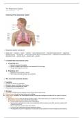
Anatomy of the respiratory system
• Respiratory system consists of:
→ Nasal cavity → pharynx → larynx → trachea → main (primary) bronchi → lobar (secondary) bronchi → segmental
(tertiary) bronchi → bronchioles → terminal bronchioles (end of conducting zone) → respiratory bronchioles (start of
respiratory zone) → alveolar ducts → alveolar sacs → alveoli
• It is divided into two functional zones:
1. Conducting zone
→ Nose to terminal bronchioles.
→ Warms, moistens, and filters air, and conducts it to the lungs.
→ No gas exchange occurs here
2. Respiratory zone
→ Respiratory bronchioles to alveoli.
→ Site of gas exchange between air and blood.
→ The nose and paranasal sinuses
• Functions:
→ Provides an airway for respiration.
→ Moistens, warms, and filters incoming air.
→ Contains olfactory receptors for smell.
→ Resonates sound during speech.
• Structure:
→ The nose is divided into the external nose and the internal nasal cavity.
► External nose: formed by nasal bones and cartilage.
► Nasal cavity: divided by the nasal septum (made of bone and cartilage) and lined with two types of mucous
membrane.
→ Olfactory mucosa: lines the slit-like superior region of the nasal cavity and contains smell receptors.
→ Respiratory mucosa: lines most of the nasal cavity.
► Contain goblet cells and seromucous nasal glands.
→ Seromucous nasal glands contain mucus-secreting cells and serous cells that secrete a watery fluid
containing enzymes.
→ Mucus traps dust, bacteria and other debris.
→ Lysozyme (an antibacterial enzyme) destroys bacteria .
→ The epithelial cells of the respiratory mucosa also secrete defensins (natural Antibiotics) that help kill
invading microbes.
→ Paranasal sinuses: surround the nasal cavity, helping lighten the skull and warm and moisten air.
→ Produce mucus.
→ Nasolacrimal ducts: drain tears from the eyes into the nasal cavity.
, → Seromucous nasal glands contain mucus-secreting cells and serous cells that secrete a watery fluid
containing enzymes.
→ Mucus traps dust, bacteria and other debris.
→ Lysozyme (an antibacterial enzyme) destroys bacteria .
→ The epithelial cells of the respiratory mucosa also secrete defensins (natural Antibiotics) that help kill
invading microbes.
→ Paranasal sinuses: surround the nasal cavity, helping lighten the skull and warm and moisten air.
→ Produce mucus.
→ Nasolacrimal ducts: drain tears from the eyes into the nasal cavity.
→ The pharynx (throat)
• A muscular tube extending from the base of the skull to C6 vertebra.
• Connects the nasal cavity and mouth to the larynx and esophagus.
• Subdivisions:
1. Nasopharynx
• Behind the nasal cavity.
• Passageway for air only.
• Continuous with the nasal cavity, taking over the job of propelling mucus.
• Contain pseudostratified ciliated epithelium.
• Contains pharyngeal tonsils (adenoids), which traps and destroys pathogens.
• During swallowing, the soft palate and its pendulous uvula move upwards, closing off the nasopharynx to
prevent food from entering the nasal cavity.
2. Oropharynx
• Behind the mouth.
• Passageway for food and air.
• Contains more protective stratified squamous epithelium to accommodate the increased friction and chemical
trauma (characteristic of hot and spicy foods).
• Contains palatine and lingual tonsils.
3. Laryngopharynx
• Above the epiglottis, extending to the larynx (air) and esophagus (food).
• Passageway for food and air.
• Contains stratified squamous epithelium.
→ The Larynx (voice box)
• Connects pharynx to trachea.
,→ The Larynx (voice box)
• Connects pharynx to trachea.
• Functions:
→ Provides a patent airway.
→ Routes food and air into proper channels (via the epiglottis).
→ Prevents food from entering lower respiratory tract.
→ Houses vocal folds/cords (acts as voice box).
• Structure:
→ Consists of cartilage and dense connective tissue
→ Opening (glottis) can be closed by epiglottis or vocal folds.
→ Cartilages:
• Thyroid cartilage
→ Formed by the fusion of two cartilage plates.
→ The midline laryngeal prominence, is obvious externally as the Adam’s apple.
• Cricoid cartilage
→ Ring shaped, below thyroid.
→ Cointain three pairs of small cartilage (arytenoid, cuneiform and corniculate cartilage).
→ Arytenoid cartilage anchor the vocal chords.
• Epiglottis
→ Flap that covers the larynx inlet during swallowing, preventing aspiration.
► Aspiration: inhaling something into the respiratory tract that should normally go into the digestive tract.
→ Composed of elastic cartilage and covered by a taste bud–containing mucosa.
→ Initiates coughing reflex when anything other than air enters respiaroty tract.
→ The Trachea (windpipe)
• Extends from the larynx to the main bronchi (around the level of T5).
• The open part of the ring faces posteriorly, allowing the esophagus to expand during swallowing.
• Supported by C-shaped rings of hyaline cartilage, which prevent collapse.
• Tracheal walls consists of a mucosa, submucosa and adventitia layer.
• The mucosa is ciliated pseudostratified columnar epithelium, which traps particles and moves them upward.
• Submucu
, → The bronchial tree
► The bronchus
• Right and left main (primary) bronchi branch from the trachea and enter the lungs at the hilum.
→ The right main bronchus is wider, shorter, and more vertical than the left.
→ It is therefore more common for foreign objects to get stuck on right side.
• Contains mucus glands.
• Each main bronchus divides into:
○ Lobar (secondary) bronchi
→ 3 on the right, 2 on the left (one per lobe).
○ Segmental (tertiary) bronchi
• The tissue composition of main bronchi are the same as trachea.
► The bronchioles
• Bronchus branch into smaller bronchi → bronchioles → terminal bronchioles.
• Terminal bronchiole contain clara (club) cells, which secrete surfactant-like fluid.
• Passages smaller than 1 mm in diameter are called bronchioles.
• No mucus glands.
• Respiratory bronchioles mark the beginning of the respiratory zone, leading to:
○ Alveolar ducts → alveolar sacs → alveoli
○ Gas exchange occurs across the respiratory membrane (alveolar and capillary walls + basement
membrane).
• Structural changes as airways get smaller:
1. Cartilage decreases
○ Irregular plates of cartilage replace the cartilage rings, and bronchiole no longer contain any cartilage.
2. Epithelium type changes.
○ It thins from pseudostratified columnar to columnar to cuboidal in terminal bronchiole.
○ Mucus-producing cells and cilia are sparse in the bronchioles.
3. Smooth muscle increases.
○ Allows bronchioles to constrict or dilate.
→ The alveoli
• Tiny, thin-walled air sacs in the lungs where gas exchange takes place.
→ Oxygen enters the blood, and carbon dioxide leaves it.
• Each alveolus is surrounded by capillaries.
• Each alveolus is surrounded by elastic fibers, allowing lungs to stretch and recoil.
• Alveolar pore: openings that connect neighboring alveoli.
→ Allows for air pressure to be equalized and provides alternate airflow routes if a bronchiole is blocked.
• The wall of an alveolus is made up of:
1. Type I alveolar cells (type I pneumocytes)
→ Simple squamous epithelial cells
→ Form major part of the alveolar wall (about 95%)
→ Very thin to allow rapid diffusion of gases.
1. Type II alveolar cells (type II pneumocytes)
→ Cuboidal epithelial cells scattered among type I cells.
→ Secrete surfactant.
→ A substance that reduces surface tension inside the alveolus so it doesn’t collapse during exhalation.
→ Also secrete antimicrobial proteins that help defend against pathogens.
1. Alveolar macrophages (dust cells)
→ Mobile immune cells that move along alveolar surfaces.
→ Phagocytose (engulf) dust, debris, and microorganisms
→ Once they’ve done their job, they’re swept up and swallowed or expelled.
• The respiratory membrane (also called the blood–air barrier) is where gas exchange occurs.
→ Diffusion is very efficient due to being very thin and having a large surface area.
Friday, 7 November 2025 15:32
Anatomy of the respiratory system
• Respiratory system consists of:
→ Nasal cavity → pharynx → larynx → trachea → main (primary) bronchi → lobar (secondary) bronchi → segmental
(tertiary) bronchi → bronchioles → terminal bronchioles (end of conducting zone) → respiratory bronchioles (start of
respiratory zone) → alveolar ducts → alveolar sacs → alveoli
• It is divided into two functional zones:
1. Conducting zone
→ Nose to terminal bronchioles.
→ Warms, moistens, and filters air, and conducts it to the lungs.
→ No gas exchange occurs here
2. Respiratory zone
→ Respiratory bronchioles to alveoli.
→ Site of gas exchange between air and blood.
→ The nose and paranasal sinuses
• Functions:
→ Provides an airway for respiration.
→ Moistens, warms, and filters incoming air.
→ Contains olfactory receptors for smell.
→ Resonates sound during speech.
• Structure:
→ The nose is divided into the external nose and the internal nasal cavity.
► External nose: formed by nasal bones and cartilage.
► Nasal cavity: divided by the nasal septum (made of bone and cartilage) and lined with two types of mucous
membrane.
→ Olfactory mucosa: lines the slit-like superior region of the nasal cavity and contains smell receptors.
→ Respiratory mucosa: lines most of the nasal cavity.
► Contain goblet cells and seromucous nasal glands.
→ Seromucous nasal glands contain mucus-secreting cells and serous cells that secrete a watery fluid
containing enzymes.
→ Mucus traps dust, bacteria and other debris.
→ Lysozyme (an antibacterial enzyme) destroys bacteria .
→ The epithelial cells of the respiratory mucosa also secrete defensins (natural Antibiotics) that help kill
invading microbes.
→ Paranasal sinuses: surround the nasal cavity, helping lighten the skull and warm and moisten air.
→ Produce mucus.
→ Nasolacrimal ducts: drain tears from the eyes into the nasal cavity.
, → Seromucous nasal glands contain mucus-secreting cells and serous cells that secrete a watery fluid
containing enzymes.
→ Mucus traps dust, bacteria and other debris.
→ Lysozyme (an antibacterial enzyme) destroys bacteria .
→ The epithelial cells of the respiratory mucosa also secrete defensins (natural Antibiotics) that help kill
invading microbes.
→ Paranasal sinuses: surround the nasal cavity, helping lighten the skull and warm and moisten air.
→ Produce mucus.
→ Nasolacrimal ducts: drain tears from the eyes into the nasal cavity.
→ The pharynx (throat)
• A muscular tube extending from the base of the skull to C6 vertebra.
• Connects the nasal cavity and mouth to the larynx and esophagus.
• Subdivisions:
1. Nasopharynx
• Behind the nasal cavity.
• Passageway for air only.
• Continuous with the nasal cavity, taking over the job of propelling mucus.
• Contain pseudostratified ciliated epithelium.
• Contains pharyngeal tonsils (adenoids), which traps and destroys pathogens.
• During swallowing, the soft palate and its pendulous uvula move upwards, closing off the nasopharynx to
prevent food from entering the nasal cavity.
2. Oropharynx
• Behind the mouth.
• Passageway for food and air.
• Contains more protective stratified squamous epithelium to accommodate the increased friction and chemical
trauma (characteristic of hot and spicy foods).
• Contains palatine and lingual tonsils.
3. Laryngopharynx
• Above the epiglottis, extending to the larynx (air) and esophagus (food).
• Passageway for food and air.
• Contains stratified squamous epithelium.
→ The Larynx (voice box)
• Connects pharynx to trachea.
,→ The Larynx (voice box)
• Connects pharynx to trachea.
• Functions:
→ Provides a patent airway.
→ Routes food and air into proper channels (via the epiglottis).
→ Prevents food from entering lower respiratory tract.
→ Houses vocal folds/cords (acts as voice box).
• Structure:
→ Consists of cartilage and dense connective tissue
→ Opening (glottis) can be closed by epiglottis or vocal folds.
→ Cartilages:
• Thyroid cartilage
→ Formed by the fusion of two cartilage plates.
→ The midline laryngeal prominence, is obvious externally as the Adam’s apple.
• Cricoid cartilage
→ Ring shaped, below thyroid.
→ Cointain three pairs of small cartilage (arytenoid, cuneiform and corniculate cartilage).
→ Arytenoid cartilage anchor the vocal chords.
• Epiglottis
→ Flap that covers the larynx inlet during swallowing, preventing aspiration.
► Aspiration: inhaling something into the respiratory tract that should normally go into the digestive tract.
→ Composed of elastic cartilage and covered by a taste bud–containing mucosa.
→ Initiates coughing reflex when anything other than air enters respiaroty tract.
→ The Trachea (windpipe)
• Extends from the larynx to the main bronchi (around the level of T5).
• The open part of the ring faces posteriorly, allowing the esophagus to expand during swallowing.
• Supported by C-shaped rings of hyaline cartilage, which prevent collapse.
• Tracheal walls consists of a mucosa, submucosa and adventitia layer.
• The mucosa is ciliated pseudostratified columnar epithelium, which traps particles and moves them upward.
• Submucu
, → The bronchial tree
► The bronchus
• Right and left main (primary) bronchi branch from the trachea and enter the lungs at the hilum.
→ The right main bronchus is wider, shorter, and more vertical than the left.
→ It is therefore more common for foreign objects to get stuck on right side.
• Contains mucus glands.
• Each main bronchus divides into:
○ Lobar (secondary) bronchi
→ 3 on the right, 2 on the left (one per lobe).
○ Segmental (tertiary) bronchi
• The tissue composition of main bronchi are the same as trachea.
► The bronchioles
• Bronchus branch into smaller bronchi → bronchioles → terminal bronchioles.
• Terminal bronchiole contain clara (club) cells, which secrete surfactant-like fluid.
• Passages smaller than 1 mm in diameter are called bronchioles.
• No mucus glands.
• Respiratory bronchioles mark the beginning of the respiratory zone, leading to:
○ Alveolar ducts → alveolar sacs → alveoli
○ Gas exchange occurs across the respiratory membrane (alveolar and capillary walls + basement
membrane).
• Structural changes as airways get smaller:
1. Cartilage decreases
○ Irregular plates of cartilage replace the cartilage rings, and bronchiole no longer contain any cartilage.
2. Epithelium type changes.
○ It thins from pseudostratified columnar to columnar to cuboidal in terminal bronchiole.
○ Mucus-producing cells and cilia are sparse in the bronchioles.
3. Smooth muscle increases.
○ Allows bronchioles to constrict or dilate.
→ The alveoli
• Tiny, thin-walled air sacs in the lungs where gas exchange takes place.
→ Oxygen enters the blood, and carbon dioxide leaves it.
• Each alveolus is surrounded by capillaries.
• Each alveolus is surrounded by elastic fibers, allowing lungs to stretch and recoil.
• Alveolar pore: openings that connect neighboring alveoli.
→ Allows for air pressure to be equalized and provides alternate airflow routes if a bronchiole is blocked.
• The wall of an alveolus is made up of:
1. Type I alveolar cells (type I pneumocytes)
→ Simple squamous epithelial cells
→ Form major part of the alveolar wall (about 95%)
→ Very thin to allow rapid diffusion of gases.
1. Type II alveolar cells (type II pneumocytes)
→ Cuboidal epithelial cells scattered among type I cells.
→ Secrete surfactant.
→ A substance that reduces surface tension inside the alveolus so it doesn’t collapse during exhalation.
→ Also secrete antimicrobial proteins that help defend against pathogens.
1. Alveolar macrophages (dust cells)
→ Mobile immune cells that move along alveolar surfaces.
→ Phagocytose (engulf) dust, debris, and microorganisms
→ Once they’ve done their job, they’re swept up and swallowed or expelled.
• The respiratory membrane (also called the blood–air barrier) is where gas exchange occurs.
→ Diffusion is very efficient due to being very thin and having a large surface area.