Brain and Cognition 1 Summary
Chapter 2 – neuroanatomy
§2.1
The brain needs sensory information (vision, audition, olfaction, gustation &
somatosensation) to produce its primary function movement (behaviour).
Agenesis: failure of brain regions to develop.
People that suffer agenesis of the cerebellum are able to function properly and only
have some problems with balance and speech. They might show symptoms of autism early in
life.
Adaptations caused by evolution have equipped each species with a view of the world that
helps them to survive.
Brain plasticity: neural tissue has the capacity to change in response to the world by
changing how it is organized. Connections between neurons are constantly changing in
response to new experiences.
Neuroplasticity: the nervous system’s potential to alter itself physically or chemically in
response to environment and to compensate for age-related changes and injury.
Phenotypic plasticity: an individual’s potential to develop a range of phenotypes (exterior
characteristics/looks)
Genotype: genetic makeup.
Epigenetic factors do not change genes, but influence which genes inherited from parents
express their traits.
Brain
Central Nervous System (CNS)
Nervous system Spinal cord
Somatic NS
Peripheral Nervous System (PNS)
Autonomic
NS
Enteric NS
Central nervous system: mediates behaviour
,Somatic nervous system (SNS): all spinal and cranial nerves. Carries sensory information to
the CNS from muscles, joints and skin. Also outgoing motor instructions that produce
movement
Autonomic nervous system (ANS): rest-and-digest response through parasympathetic
(calming) nerves and fight-or-flight response through sympathetic (arousing) nerves.
Enteric nervous system (ENS): neurons in the lining of the gut that control the gut. Mostly
operates autonomously, but can communicate with the CNS through the ANS.
Afferent = incoming
Efferent = outgoing (E > exit)
The first thing you encounter after opening the skull is not the brain, but Meninges. This is a
triple layered protection that consists of the outer dura mater, middle arachnoid layer and
inner pia mater.
Between the pia mater and arachnoid layer flows cerebrospinal fluid (CSF). This is a
colourless solution of sodium (na+), chloride (cl-) and other ions. Its function is to allow the
brain to expand slightly without pressing on the skull.
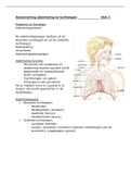
Outer layer of the brain = cerebral cortex. It is heavily folded and layered to fit inside the
brain.
Lobes of the brain:
1. Frontal lobe: performs brain’s executive functions, like decision making and voluntary
movement.
2. Parietal lobe: directing movement towards a goal or to perform a task.
3. Temporal lobe: functions include hearing, language, musical abilities, facial
recognition and emotional processing.
4. Occipital lobe: start of visual scene processing
,Brainstem: heart rate, breathing, sleeping and
eating.
Meningitis: inflammation of the meninges. The
increase in white blood cells by the body
increases cranial pressure which can lead to
delirium and if the infection progresses can lead
to drowsiness, coma, stupor and even death.
Encephalitis: infection of the brain itself.
Caused by viruses.
Bumps: Gyri (Gyrus)
Cracks: Sulci (Sulcus)
Really deep sulci: Fissures
Three major arteries send blood to the
cerebrum: Anterior, Middle and Posterior
Cerebral arteries.
Dark, outer regions of brain:
Gray matter. This is largely
composed of cell bodies and
capillary blood vessels. Neurons collect and modify information to send it along here.
Light, inner regions of the brain: White matter. Mostly nerve fibers covered in myelin
sheaths that have high fat content. This is what makes the matter appear to be white. White
matter forms long-distance connections between neurons.
There are 4 ventricles in total.
, Lateral ventricles: two wing-shaped cavities filled with cerebrospinal fluid.
All ventricles contain CSF. They eventually run into the cerebral aqueduct. This is a canal
that runs down the length of the spinal cord.
CSF performs several vital brain functions.
- Naturally buoyant, so it appears to weigh only about 1/30 of its actual mass
- Acts as a shock absorber.
The CSF environment is carefully made up. Small changes in the chemical content can result
in dizziness and fainting.
Corpus collosum: contains about 200 million nerve fibers that join the left and right
hemispheres and allows them to communicate.
Subcortical regions: regions below (inside) the neocortex.
Neurons: carry out the brain’s communicative and information processing functions.
Glial cells: aid the neurons
Nuclei: a cluster of similar cells
Tract: collection of nerve fibers in the brain and spinal cord
Nerve: bundles of fibers outside the CNS.
Chapter 2 – neuroanatomy
§2.1
The brain needs sensory information (vision, audition, olfaction, gustation &
somatosensation) to produce its primary function movement (behaviour).
Agenesis: failure of brain regions to develop.
People that suffer agenesis of the cerebellum are able to function properly and only
have some problems with balance and speech. They might show symptoms of autism early in
life.
Adaptations caused by evolution have equipped each species with a view of the world that
helps them to survive.
Brain plasticity: neural tissue has the capacity to change in response to the world by
changing how it is organized. Connections between neurons are constantly changing in
response to new experiences.
Neuroplasticity: the nervous system’s potential to alter itself physically or chemically in
response to environment and to compensate for age-related changes and injury.
Phenotypic plasticity: an individual’s potential to develop a range of phenotypes (exterior
characteristics/looks)
Genotype: genetic makeup.
Epigenetic factors do not change genes, but influence which genes inherited from parents
express their traits.
Brain
Central Nervous System (CNS)
Nervous system Spinal cord
Somatic NS
Peripheral Nervous System (PNS)
Autonomic
NS
Enteric NS
Central nervous system: mediates behaviour
,Somatic nervous system (SNS): all spinal and cranial nerves. Carries sensory information to
the CNS from muscles, joints and skin. Also outgoing motor instructions that produce
movement
Autonomic nervous system (ANS): rest-and-digest response through parasympathetic
(calming) nerves and fight-or-flight response through sympathetic (arousing) nerves.
Enteric nervous system (ENS): neurons in the lining of the gut that control the gut. Mostly
operates autonomously, but can communicate with the CNS through the ANS.
Afferent = incoming
Efferent = outgoing (E > exit)
The first thing you encounter after opening the skull is not the brain, but Meninges. This is a
triple layered protection that consists of the outer dura mater, middle arachnoid layer and
inner pia mater.
Between the pia mater and arachnoid layer flows cerebrospinal fluid (CSF). This is a
colourless solution of sodium (na+), chloride (cl-) and other ions. Its function is to allow the
brain to expand slightly without pressing on the skull.
Outer layer of the brain = cerebral cortex. It is heavily folded and layered to fit inside the
brain.
Lobes of the brain:
1. Frontal lobe: performs brain’s executive functions, like decision making and voluntary
movement.
2. Parietal lobe: directing movement towards a goal or to perform a task.
3. Temporal lobe: functions include hearing, language, musical abilities, facial
recognition and emotional processing.
4. Occipital lobe: start of visual scene processing
,Brainstem: heart rate, breathing, sleeping and
eating.
Meningitis: inflammation of the meninges. The
increase in white blood cells by the body
increases cranial pressure which can lead to
delirium and if the infection progresses can lead
to drowsiness, coma, stupor and even death.
Encephalitis: infection of the brain itself.
Caused by viruses.
Bumps: Gyri (Gyrus)
Cracks: Sulci (Sulcus)
Really deep sulci: Fissures
Three major arteries send blood to the
cerebrum: Anterior, Middle and Posterior
Cerebral arteries.
Dark, outer regions of brain:
Gray matter. This is largely
composed of cell bodies and
capillary blood vessels. Neurons collect and modify information to send it along here.
Light, inner regions of the brain: White matter. Mostly nerve fibers covered in myelin
sheaths that have high fat content. This is what makes the matter appear to be white. White
matter forms long-distance connections between neurons.
There are 4 ventricles in total.
, Lateral ventricles: two wing-shaped cavities filled with cerebrospinal fluid.
All ventricles contain CSF. They eventually run into the cerebral aqueduct. This is a canal
that runs down the length of the spinal cord.
CSF performs several vital brain functions.
- Naturally buoyant, so it appears to weigh only about 1/30 of its actual mass
- Acts as a shock absorber.
The CSF environment is carefully made up. Small changes in the chemical content can result
in dizziness and fainting.
Corpus collosum: contains about 200 million nerve fibers that join the left and right
hemispheres and allows them to communicate.
Subcortical regions: regions below (inside) the neocortex.
Neurons: carry out the brain’s communicative and information processing functions.
Glial cells: aid the neurons
Nuclei: a cluster of similar cells
Tract: collection of nerve fibers in the brain and spinal cord
Nerve: bundles of fibers outside the CNS.