What brain regions are involved in the types of movement?
Primary motor cortex: responsible for execution of all voluntary movements of the body
different regions of the primary motor cortex represent different regions of the body =
somatotopically organized
- The left hemisphere is specialized for movements on the right side of the body
- The right hemisphere is specialized for movements on the left side of the body
The activity for each neuron is highest for a particular direction of movement (the
preferred direction) and it decreases gradually with directions further and further away
Bulboreticular facilitatory region of the brain stem (secondarily by impulses transmitted
from the cerebellum, basal ganglia and cerebral cortex)
When a person must perform a muscle function that requires high degree of delicate
and exact positioning excitation of the appropriate muscle spindles by signals from
the bulboreticular facilitatory region stabilizes the position of the major joints
3 types of movements:
1. Reflex movements
= Mostly spinal cord 2 types of reflex movements:
- Postural reflexes
-
2. Voluntary movements cerebral cortex
3. Rhythmic movements combination of reflex and voluntary control.
Motor cortex
3 parts: premotor cortex, supplementary cortex cingulate cortex area (links emotion to
movements)
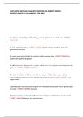
, What is the anatomy of the spinal cord?
The gray matter is the integrative area for the spinal cord reflexes
Sensory signals enter the cord almost entirely through the dorsal root. After entering the
spinal cord, every sensory signal travels to 2 separate destinations:
1) One branch of the sensory nerve
terminates almost immediately in the
gray matter and elicits local segmental
SC reflexes and other local effects
2) Another branch transmits signals to
higher levels of NS (brainstem, cerebral
cortex)
Different types of neurons:
- Sensory relay neurons
- Anterior motor neurons
- Interneurons
Central canal: centre of SC, CSF in it
2 types of Lower motor neurons:
Anterior motor neurons:
= located in each segment of the anterior horns of SC gray matter. Are larger than most
of the other neurons
- Give rise to nerve fibers that leave the SC by way of anterior roots and directly
innervate the skeletal muscle fibers
2 types of anterior motor neurons:
1. Alpha motor neurons
Give rise to large type A alpha motor nerve fibers. They branch many times after they
enter the muscle and innervate the large skeletal muscle fibers. Innervate the
extrafusal skeletal muscle
Stimulation of 1 single alpha nerve fiber excites anywhere from 3 to several 100
skeletal muscle fibers = collectively called motor unit
Fast fatigue resistance, slow, type I (slow), type II has 2 types
2. Gamma motor neurons
Primary motor cortex: responsible for execution of all voluntary movements of the body
different regions of the primary motor cortex represent different regions of the body =
somatotopically organized
- The left hemisphere is specialized for movements on the right side of the body
- The right hemisphere is specialized for movements on the left side of the body
The activity for each neuron is highest for a particular direction of movement (the
preferred direction) and it decreases gradually with directions further and further away
Bulboreticular facilitatory region of the brain stem (secondarily by impulses transmitted
from the cerebellum, basal ganglia and cerebral cortex)
When a person must perform a muscle function that requires high degree of delicate
and exact positioning excitation of the appropriate muscle spindles by signals from
the bulboreticular facilitatory region stabilizes the position of the major joints
3 types of movements:
1. Reflex movements
= Mostly spinal cord 2 types of reflex movements:
- Postural reflexes
-
2. Voluntary movements cerebral cortex
3. Rhythmic movements combination of reflex and voluntary control.
Motor cortex
3 parts: premotor cortex, supplementary cortex cingulate cortex area (links emotion to
movements)
, What is the anatomy of the spinal cord?
The gray matter is the integrative area for the spinal cord reflexes
Sensory signals enter the cord almost entirely through the dorsal root. After entering the
spinal cord, every sensory signal travels to 2 separate destinations:
1) One branch of the sensory nerve
terminates almost immediately in the
gray matter and elicits local segmental
SC reflexes and other local effects
2) Another branch transmits signals to
higher levels of NS (brainstem, cerebral
cortex)
Different types of neurons:
- Sensory relay neurons
- Anterior motor neurons
- Interneurons
Central canal: centre of SC, CSF in it
2 types of Lower motor neurons:
Anterior motor neurons:
= located in each segment of the anterior horns of SC gray matter. Are larger than most
of the other neurons
- Give rise to nerve fibers that leave the SC by way of anterior roots and directly
innervate the skeletal muscle fibers
2 types of anterior motor neurons:
1. Alpha motor neurons
Give rise to large type A alpha motor nerve fibers. They branch many times after they
enter the muscle and innervate the large skeletal muscle fibers. Innervate the
extrafusal skeletal muscle
Stimulation of 1 single alpha nerve fiber excites anywhere from 3 to several 100
skeletal muscle fibers = collectively called motor unit
Fast fatigue resistance, slow, type I (slow), type II has 2 types
2. Gamma motor neurons