

Respiratory System
Respiratory Structures
cells use oxygen and produce carbon dioxide
the RS (Respiratory System) brings the needed oxygen into and eliminates CO2 from the body
blood transports these gases
carrying O2 to tissues
carrying CO2 to lungs
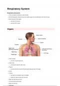
Organs
nares (nostrils)
air enters nose through here
Nasal cavity
air passes through this after nares
Pharynx
muscular tube that carries both food and air through most of its length
Larynx
Trachea
held open by incomplete rings of cartilage
divides into right and left main bronchus
Right main (primary) bronchus
Left main (primary) bronchus
Right and Left lung
Diaphragm
, each lung is surrounded y 2 layers of serous membrane aka pleurae
the relationship between pleurae and lungs can be demonstrated by pushing a fist into a water filled
balloon
balloon - pleurae
fist - lung
the balloon wraps around the fist when pushed
opposite surfaces of the balloon almost touch
inner part of balloon that wraps around fist - visceral pleura
outer part of balloon - parietal pleura
water filled space between two layers - pleural cavity
the visceral and parietal pleurae are actually a continuation of the same membrane
visceral pleura parietal pleura pleural cavity
part of pleurae lines contains pleural fluid
covers surface of lungs mediastinum
diaphragm
thoracic wall
Visceral and Parietal Pleurae
enclose each lung in a separate sac
parietal pleurae
lines the interior thoracic wall
lines the mediastinum, superior surface of
Respiratory Structures
cells use oxygen and produce carbon dioxide
the RS (Respiratory System) brings the needed oxygen into and eliminates CO2 from the body
blood transports these gases
carrying O2 to tissues
carrying CO2 to lungs
Organs
nares (nostrils)
air enters nose through here
Nasal cavity
air passes through this after nares
Pharynx
muscular tube that carries both food and air through most of its length
Larynx
Trachea
held open by incomplete rings of cartilage
divides into right and left main bronchus
Right main (primary) bronchus
Left main (primary) bronchus
Right and Left lung
Diaphragm
, each lung is surrounded y 2 layers of serous membrane aka pleurae
the relationship between pleurae and lungs can be demonstrated by pushing a fist into a water filled
balloon
balloon - pleurae
fist - lung
the balloon wraps around the fist when pushed
opposite surfaces of the balloon almost touch
inner part of balloon that wraps around fist - visceral pleura
outer part of balloon - parietal pleura
water filled space between two layers - pleural cavity
the visceral and parietal pleurae are actually a continuation of the same membrane
visceral pleura parietal pleura pleural cavity
part of pleurae lines contains pleural fluid
covers surface of lungs mediastinum
diaphragm
thoracic wall
Visceral and Parietal Pleurae
enclose each lung in a separate sac
parietal pleurae
lines the interior thoracic wall
lines the mediastinum, superior surface of


