Week 1: Cardiovascular
1. Review the normal anatomy and physiology of the cardiovascular
system.
• Structures and Functions of the Cardiovascular System
o Heart
▪ Structure
▪ Blood flow through the heart
• Cardiac valves
▪ Blood supply to the myocardium
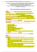
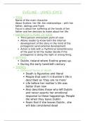
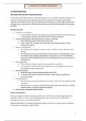
o FIG. 34.1 Schematic representation of blood flow
through the heart. Arrows indicate direction of flow. 1,
The right atrium receives venous blood from the inferior
and superior venae cavae and the coronary
sinus. The blood then passes through the
tricuspid valve into the right ventricle. 2, With
each contraction, the right ventricle pumps
blood through the pulmonic valve into the
pulmonary artery and to the lungs. 3,
Oxygenated blood flows from the lungs to the
left atrium by way of the pulmonary veins. 4, It
then passes through the mitral valve and into
the left ventricle. 5, As the heart contracts, blood
is ejected through the aortic valve into the aorta
and thus enters the systemic circulation.
o Anatomical structures of the heart →
o Coronary arteries and veins →
• Conduction system
o Cardiac cycle
o Electrocardiogram (P, QRS, T, U waveforms)
• Mechanical system
o Systole, diastole, cardiac output
• Factors affecting cardiac output
1
, o Preload, contractility, afterload
• Cardiac reserve
o Increasing cardiac output
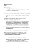
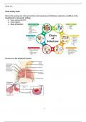
• FIG. 34.4 A, Conduction system of the heart. AV, atrioventricular; SA, sinoatrial. B, The
normal electrocardiogram pattern. The P wave represents depolarization of the atria.
The QRS complex indicates depolarization of the ventricles. The T wave represents
repolarization of the ventricles. The U wave, if present, may represent repolarization of
the Purkinje fibres or may be associated with hypokalemia. The PR, QRS, and QT
intervals reflect the length of time it takes for the impulse to travel from one area of the
heart to another.
• Vascular system
o Blood vessels
o Arteries, arterioles
▪ Capillaries
▪ Veins and venules
o Comparative thickness of layers of an artery, a vein, and a
capillary →
• Regulation of the cardiovascular system
o Autonomic nervous system
▪ Effects on the heart
▪ Effects on the blood vessels
o Baroreceptors
o Chemoreceptors
• Blood pressure
o Measurement of arterial blood pressure
o Pulse pressure and mean arterial pressure
Cardiovascular Review
• Structure: four chamber organ that lies between the lungs and the thorax
• Layers: Heart has 3 layers”
o (1) Endocardium: the inner lining, continuous with the heart valves.
o (2) Myocardium: middle, muscle layers
o (3) Epicardium: fibrous, outer layer
2
, ▪ Heart is surrounded by the Pericardium, which is a sac that holds the heart in
place and protects it.
▪ The inner serous layer.
▪ The outer fibrous layer.
▪ The fluid that exists in this space is called the serous fluid.
• Heart Divided: is divided by the septum. Has a left side
and a right side. They do not exchange blood between
the two sides.
• The atria are filling chambers and
• The ventricles are pumping chambers
The heart is also divided into two (2) circuits :
• Pulmonary circuit- pumps blood to the lungs. Which side
of the heart makes up the pulmonary circuit?
• Systemic circuit- supplies oxygenated blood and
nutrients to the entire body. Which side of the heart
makes up the systemic circuit?
Coronary Circulation
o Cardiac muscle fibers (cardiac cells) & the other types of cells in the myocardium
are not nourished by the blood in the heart chambers.
o These cells receive nutrients and rid themselves of wastes at capillaries embedded in the heart
wall - called the coronary circulation.
o There are 2 coronary arteries - right & left - which branches from the aorta just superior to the
aortic semilunar valve.
Conduction System
o The conduction system is a specialized group of cardiac muscle fibers that initiate & stimulate
contraction of the atria and ventricle.
o It is said to be intrinsic - meaning the heart beats automatically without the need for external
nervous stimulation.
o Remember, without the conduction system, the atria & ventricles would contract at different
rates.
Cardiovascular Concept Review
• Vasodilation of veins would result in decrease preload.
• Arterial vasoconstriction would result in increase in afterload.
• A slight increase in the heart rate as compensation would increase cardiac output.
• An increase in afterload can result in decrease cardiac output.
• Drugs that increase myocardial contractility are positive inotropes.
• Drugs that decrease myocardial contractility are negative inotropes.
• Drugs that increase heart rate are positive chronotropes.
3
, • Drugs that decrease heart rate are negative chronotropes.
2. Discuss arteriosclerosis and atherosclerosis under the following headings:
• Etiology
o Atherosclerosis is the major cause of coronary artery
disease.
▪ It is characterized by deposits of lipids within the
intima of the artery.
▪ Endothelial injury and inflammation play a central
role in the development of atherosclerosis.
• Pathophysiology
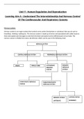
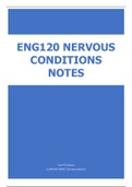
o FIG. 36.1 Pathogenesis of atherosclerosis. A, Damaged
endothelium. B, Fatty streak and lipid core formation. C,
Fibrous plaque. Raised plaques are visible: some are yellow;
others are white. D, Complicated lesion: thrombus is red;
collagen is blue. Plaque is complicated by red thrombus
deposition.
o Developmental Stages
▪ Fatty streak
▪ Fibrous plaque
▪ Complicated lesion
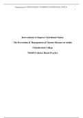
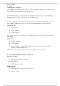
o Collateral Circulation
▪ It is an alternate or “backup” blood vessels in your
body that can take over when another artery or
vein becomes blocked or damaged. Your collateral
circulation provides alternative
routes for blood flow.
▪ FIG. 36.2 Vessel occlusion with
collateral circulation. A, Open,
functioning coronary artery. B,
Partial coronary artery closure with
collateral circulation being
established. C, Total coronary
artery occlusion with collateral
circulation bypassing the occlusion
to supply the myocardium
• Risk Factors for Coronary Artery Disease
o Nonmodifiable risk factors
▪ Age, sex, and ethnicity
▪ Family history and genetics
o Modifiable major risk factors
▪ Elevated serum lipid levels
▪ Elevated blood pressure (BP)
▪ Tobacco use
▪ Physical inactivity
▪ Obesity
▪ Diabetes mellitus
▪ Elevated fasting bold glucose level
4
1. Review the normal anatomy and physiology of the cardiovascular
system.
• Structures and Functions of the Cardiovascular System
o Heart
▪ Structure
▪ Blood flow through the heart
• Cardiac valves
▪ Blood supply to the myocardium
o FIG. 34.1 Schematic representation of blood flow
through the heart. Arrows indicate direction of flow. 1,
The right atrium receives venous blood from the inferior
and superior venae cavae and the coronary
sinus. The blood then passes through the
tricuspid valve into the right ventricle. 2, With
each contraction, the right ventricle pumps
blood through the pulmonic valve into the
pulmonary artery and to the lungs. 3,
Oxygenated blood flows from the lungs to the
left atrium by way of the pulmonary veins. 4, It
then passes through the mitral valve and into
the left ventricle. 5, As the heart contracts, blood
is ejected through the aortic valve into the aorta
and thus enters the systemic circulation.
o Anatomical structures of the heart →
o Coronary arteries and veins →
• Conduction system
o Cardiac cycle
o Electrocardiogram (P, QRS, T, U waveforms)
• Mechanical system
o Systole, diastole, cardiac output
• Factors affecting cardiac output
1
, o Preload, contractility, afterload
• Cardiac reserve
o Increasing cardiac output
• FIG. 34.4 A, Conduction system of the heart. AV, atrioventricular; SA, sinoatrial. B, The
normal electrocardiogram pattern. The P wave represents depolarization of the atria.
The QRS complex indicates depolarization of the ventricles. The T wave represents
repolarization of the ventricles. The U wave, if present, may represent repolarization of
the Purkinje fibres or may be associated with hypokalemia. The PR, QRS, and QT
intervals reflect the length of time it takes for the impulse to travel from one area of the
heart to another.
• Vascular system
o Blood vessels
o Arteries, arterioles
▪ Capillaries
▪ Veins and venules
o Comparative thickness of layers of an artery, a vein, and a
capillary →
• Regulation of the cardiovascular system
o Autonomic nervous system
▪ Effects on the heart
▪ Effects on the blood vessels
o Baroreceptors
o Chemoreceptors
• Blood pressure
o Measurement of arterial blood pressure
o Pulse pressure and mean arterial pressure
Cardiovascular Review
• Structure: four chamber organ that lies between the lungs and the thorax
• Layers: Heart has 3 layers”
o (1) Endocardium: the inner lining, continuous with the heart valves.
o (2) Myocardium: middle, muscle layers
o (3) Epicardium: fibrous, outer layer
2
, ▪ Heart is surrounded by the Pericardium, which is a sac that holds the heart in
place and protects it.
▪ The inner serous layer.
▪ The outer fibrous layer.
▪ The fluid that exists in this space is called the serous fluid.
• Heart Divided: is divided by the septum. Has a left side
and a right side. They do not exchange blood between
the two sides.
• The atria are filling chambers and
• The ventricles are pumping chambers
The heart is also divided into two (2) circuits :
• Pulmonary circuit- pumps blood to the lungs. Which side
of the heart makes up the pulmonary circuit?
• Systemic circuit- supplies oxygenated blood and
nutrients to the entire body. Which side of the heart
makes up the systemic circuit?
Coronary Circulation
o Cardiac muscle fibers (cardiac cells) & the other types of cells in the myocardium
are not nourished by the blood in the heart chambers.
o These cells receive nutrients and rid themselves of wastes at capillaries embedded in the heart
wall - called the coronary circulation.
o There are 2 coronary arteries - right & left - which branches from the aorta just superior to the
aortic semilunar valve.
Conduction System
o The conduction system is a specialized group of cardiac muscle fibers that initiate & stimulate
contraction of the atria and ventricle.
o It is said to be intrinsic - meaning the heart beats automatically without the need for external
nervous stimulation.
o Remember, without the conduction system, the atria & ventricles would contract at different
rates.
Cardiovascular Concept Review
• Vasodilation of veins would result in decrease preload.
• Arterial vasoconstriction would result in increase in afterload.
• A slight increase in the heart rate as compensation would increase cardiac output.
• An increase in afterload can result in decrease cardiac output.
• Drugs that increase myocardial contractility are positive inotropes.
• Drugs that decrease myocardial contractility are negative inotropes.
• Drugs that increase heart rate are positive chronotropes.
3
, • Drugs that decrease heart rate are negative chronotropes.
2. Discuss arteriosclerosis and atherosclerosis under the following headings:
• Etiology
o Atherosclerosis is the major cause of coronary artery
disease.
▪ It is characterized by deposits of lipids within the
intima of the artery.
▪ Endothelial injury and inflammation play a central
role in the development of atherosclerosis.
• Pathophysiology
o FIG. 36.1 Pathogenesis of atherosclerosis. A, Damaged
endothelium. B, Fatty streak and lipid core formation. C,
Fibrous plaque. Raised plaques are visible: some are yellow;
others are white. D, Complicated lesion: thrombus is red;
collagen is blue. Plaque is complicated by red thrombus
deposition.
o Developmental Stages
▪ Fatty streak
▪ Fibrous plaque
▪ Complicated lesion
o Collateral Circulation
▪ It is an alternate or “backup” blood vessels in your
body that can take over when another artery or
vein becomes blocked or damaged. Your collateral
circulation provides alternative
routes for blood flow.
▪ FIG. 36.2 Vessel occlusion with
collateral circulation. A, Open,
functioning coronary artery. B,
Partial coronary artery closure with
collateral circulation being
established. C, Total coronary
artery occlusion with collateral
circulation bypassing the occlusion
to supply the myocardium
• Risk Factors for Coronary Artery Disease
o Nonmodifiable risk factors
▪ Age, sex, and ethnicity
▪ Family history and genetics
o Modifiable major risk factors
▪ Elevated serum lipid levels
▪ Elevated blood pressure (BP)
▪ Tobacco use
▪ Physical inactivity
▪ Obesity
▪ Diabetes mellitus
▪ Elevated fasting bold glucose level
4