• Receptors
- Types of receptors:
➢ Photoreceptors > Receptors sensitive to light. E.g., in the retina of the eye.
➢ Mechanoreceptors > receptors sensitive to pressure changes. E.g., in the skin.
➢ Proprioceptors > receptors that respond to position and movement E.g., in the muscle.
➢ Chemoreceptors > receptors sensitive to chemical substances. E.g., taste buds.
➢ Thermoreceptors > receptors sensitive to temperature changes.
➢ Nociceptors > Receptors sensitive to pain.
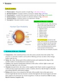
Structure of the eye – external.
- The eyeball is found within the bony
orbit of the skull cushioned by fat.
- 6 muscles control eye movement:
1. Superior rectus.
2. Inferior rectus
3. Medial rectus
4. Lateral rectus.
5. Superior oblique.
6. Inferior oblique.
- The lacrimal gland, situated at the
outer corner of the eye, secretes tears,
which are emptied into the lacrimal
ducts in the inner corner of the eye.
- Tears and blinking keep the surface
moist and prevent desiccation.
- Tears also have an antiseptic property.
• Structure of the eye – functions.
➢ Conjunctiva: a thin membrane that covers the cornea and also lines the eyelids. This
structure is extremely sensitive and results in blinking and tear secretion when foreign
bodies enter the eye.
➢ Sclera: the white, fibrous part of the eyeball, protects and maintains the shape of the
eyeball; it allows for attachment of ocular muscles.
➢ Cornea: is the anterior transparent extension of the sclera; it allows light to enter and
refracts the light due to its curvature.
➢ Choroid: situated below the sclera; richly supplied with blood vessels to supply the eye
with food and oxygen and remove wastes; is darkly colored so that the light is absorbed
preventing reflection and blurred images.
➢ Retina: inner layer of eye situated at the back of the eyeball, close to the optic nerve:
receives light that the lens has focused, converts the light into the neural signals and send
these signals on to the brain for visual recognition. 3 Cells are found here: Rods & cones,
Bipolar neurons, Ganglion cells.
, 1. Rods and cones:
➢ Rods: enables black & white vision and vision in lowlight conditions.
➢ Cones: enables color vision & vision in bright light conditions.
2. Bipolar neurons:
➢ Specialized sensory neurons for 5 senses.
3. Ganglion cells:
➢ Receives information from rods & cones.
Iris:
- Colored extension of cornea
- Contains pigment.
- Circular and radial muscles form diaphragm.
- Muscles are under automatic control.
- Muscles regulates size of the pupil.
- Blood vessels are also present.
Pupil:
- Situated at the center of the iris.
- Allows light to enter eye and reach retina.
- Size of pupil is controlled by muscles of Iris.
Lens:
- A biconvex, transparent structure.
- Held in place by suspensory ligaments attached to ciliary muscles.
- Lens change shape, depending on whether the object is being viewed is near
or far.
- Lens focuses light onto the retina.
Ciliary body:
- Contains ciliary muscle.
- Adjusts lens during accommodation.
- Secretes aqueous humour.
Suspensory ligaments:
- Inelastic structures that are attached to the ciliary muscles.
- Attached to the lens at the other end.
- Types of receptors:
➢ Photoreceptors > Receptors sensitive to light. E.g., in the retina of the eye.
➢ Mechanoreceptors > receptors sensitive to pressure changes. E.g., in the skin.
➢ Proprioceptors > receptors that respond to position and movement E.g., in the muscle.
➢ Chemoreceptors > receptors sensitive to chemical substances. E.g., taste buds.
➢ Thermoreceptors > receptors sensitive to temperature changes.
➢ Nociceptors > Receptors sensitive to pain.
Structure of the eye – external.
- The eyeball is found within the bony
orbit of the skull cushioned by fat.
- 6 muscles control eye movement:
1. Superior rectus.
2. Inferior rectus
3. Medial rectus
4. Lateral rectus.
5. Superior oblique.
6. Inferior oblique.
- The lacrimal gland, situated at the
outer corner of the eye, secretes tears,
which are emptied into the lacrimal
ducts in the inner corner of the eye.
- Tears and blinking keep the surface
moist and prevent desiccation.
- Tears also have an antiseptic property.
• Structure of the eye – functions.
➢ Conjunctiva: a thin membrane that covers the cornea and also lines the eyelids. This
structure is extremely sensitive and results in blinking and tear secretion when foreign
bodies enter the eye.
➢ Sclera: the white, fibrous part of the eyeball, protects and maintains the shape of the
eyeball; it allows for attachment of ocular muscles.
➢ Cornea: is the anterior transparent extension of the sclera; it allows light to enter and
refracts the light due to its curvature.
➢ Choroid: situated below the sclera; richly supplied with blood vessels to supply the eye
with food and oxygen and remove wastes; is darkly colored so that the light is absorbed
preventing reflection and blurred images.
➢ Retina: inner layer of eye situated at the back of the eyeball, close to the optic nerve:
receives light that the lens has focused, converts the light into the neural signals and send
these signals on to the brain for visual recognition. 3 Cells are found here: Rods & cones,
Bipolar neurons, Ganglion cells.
, 1. Rods and cones:
➢ Rods: enables black & white vision and vision in lowlight conditions.
➢ Cones: enables color vision & vision in bright light conditions.
2. Bipolar neurons:
➢ Specialized sensory neurons for 5 senses.
3. Ganglion cells:
➢ Receives information from rods & cones.
Iris:
- Colored extension of cornea
- Contains pigment.
- Circular and radial muscles form diaphragm.
- Muscles are under automatic control.
- Muscles regulates size of the pupil.
- Blood vessels are also present.
Pupil:
- Situated at the center of the iris.
- Allows light to enter eye and reach retina.
- Size of pupil is controlled by muscles of Iris.
Lens:
- A biconvex, transparent structure.
- Held in place by suspensory ligaments attached to ciliary muscles.
- Lens change shape, depending on whether the object is being viewed is near
or far.
- Lens focuses light onto the retina.
Ciliary body:
- Contains ciliary muscle.
- Adjusts lens during accommodation.
- Secretes aqueous humour.
Suspensory ligaments:
- Inelastic structures that are attached to the ciliary muscles.
- Attached to the lens at the other end.