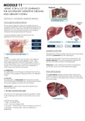
MODULE 11
‘URINE’ FOR A LOT OF LEARNING!
THE ACCESSORY DIGESTIVE ORGANS
AND URINARY SYSTEM
SECTION 01: ACCESSORY DIGESTIVE ORGANS
THE ACCESSORY DIGESTIVE ORGANS
The accessory digestive organs are a significant part of
acquiring nutrients from food. They provide enzymes for the
breakdown of food molecules and bile for the digestion of
dietary fat. This is an important process in the storage of
molecules that provide energy for the body.
Accessory digestive organs include the liver, the
gallbladder, and the pancreas.
LIGAMENTS OF THE LIVER
The ligaments of the liver attach the liver to the surrounding
abdominal peritoneum and the diaphragm.
1. Liver
The liver is an organ with many functions in the digestive The right and left lobes of the liver are separated by the
system. It produces bile for the digestion of fats. It also falciform ligament. The coronary ligament suspends the
stores dietary glucose in the form of glycogen, so that it liver from the inferior surface of the diaphragm.
can be later broken down and used for the production of
energy. The liver also plays a role in the metabolism of Peritoneum: A thin membrane that lines the abdominal
toxins, drugs, and alcohol in the blood. cavity and covers most of the abdominal organs.
2. Gallbladder
The gallbladder is a small organ underneath the liver that PORTA HEPATIS (HILUM)
functions in the storage and release of bile in the digestive
system. The porta hepatis (hepatic portal) or hilum of the liver is
where the hepatic vessels and ducts enter and leave the
3. Pancreas liver. It is located on the inferior side of the liver, surrounded
The pancreas is a mixed gland with endocrine functions by the four lobes.
that control levels of blood glucose, and exocrine functions
that secrete digestive enzymes into the intestine.
Mixed Gland: A gland which has both endocrine and
exocrine functions.
1. THE LIVER: LOCATION AND LOBES
The liver sits in the upper right abdominal quadrant, inferior
to the diaphragm and anterior to the inferior vena cava (I
V C).
The liver has four lobes:
1. Right – the largest lobe
2. Left – the second largest lobe
3. Caudate – small liver lobe, which sits adjacent to the IVC
4. Quadrate – small liver lobe, which sits adjacent to
gallbladder
, STRUCTURES OF THE PORTA HEPATIS (HILUM) PORTAL (HEPATIC) TRIADS
There are three structures that enter and leave the porta The portal triads are branches of the hepatic artery, portal
hepatis. vein, and common hepatic duct from the porta hepatis.
Within the liver there are many triads, as each lobule is
surrounded by six triads, one at each corner of the
hexagon.
a. Common Hepatic Duct
The common hepatic duct drains bile produced in the liver.
It joins with the cystic duct of the gallbladder to form the
common bile duct. These structures will be covered later in
this module when you learn about the gallbladder.
b. Portal Vein
The portal vein carries nutrient rich blood from the digestive
system into the liver, where those nutrients absorbed from
the digested food can be stored. If any toxins or drugs are
ingested, they travel through this vessel into the liver to be
metabolized.
c. Hepatic Artery
The hepatic artery carries oxygenated blood to the liver
and branches to supply each lobe.
LIVER HISTOLOGY: HEPATOCYTES LIVER LOBULE: FLOW OF VENOUS BLOOD
The functional unit of the liver is the hexagonal-shaped liver Nutrient rich blood from the portal veins travels into the
lobule. Each lobule is made up of simple cuboidal liver sinusoids. In the sinusoids, the nutrients from the blood are
cells, also known as hepatocytes, arranged in plates taken up into the hepatocytes.
(cords) that radiate outward from a central vein. Between
the plates of cells are spaces called sinusoids where venous Then, the blood in the sinusoids drains into the central veins,
blood flows. which join to form the hepatic veins. Finally, blood from
each hepatic vein drains to the inferior vena cava and
eventually to the heart.