Summary biomolecular architectures
,Chapter 1 Cytoskeleton: microfilaments
This chapter surrounds the question how diversity of cellular shape, organisation and motility is
achieved.
The cytoskeleton
Cells must have different protein combinations in order to carry out their specific functions. Often
cells have functionally distinct regions, a phenomenon that the cell achieves by cell polarity.
• the cell’s functional polarity and internal organisation are provided by the cytoskeleton: a 3D
filamentous protein network
− the cytoskeleton is attached to the plasma membrane and internal organelles →
framework for cellular organisation
− the cytoskeleton is very dynamic → reorganisation: lengths and dynamics + local
regulation
The cytoskeleton provides support and organisation. It is composed of three major filament systems.
each system is a polymer of assembled subunits → subunits regulated in time and space.
• Microfilaments: actin polymers → organised by actin binding proteins
− important in plasma membrane organisation: shape surface structures
− MFs can serve on their own or be tracks for ATP-powered myosin motor proteins →
contraction/cargo transport
• Microtubules: tubulin polymers → organised by MT-associated proteins
− provide organisational framework for cell organelles and structural support
− mitotic spindle formation
− tracks for ATP-driven cargo transport via kinesins
• Intermediate filaments: tissue-specific filamentous structures → serve many different
functions
− barrier functions, nuclear structure support
− not used as tracks by motor proteins
Cytoskeletal arrangements very between cell types → accomplished by cellular signalling for
organisation: structure determines function
• signalling may be from the ECM → signal transduction via receptors
Microfilaments and actin structures
Due to the structure-function relationship and regulated structure, microfilaments have lots of
structures that all underly particular cellular functions (via actin)
• structural role
• harnessing power of actin polymerisation to do work
• tracks for myosin motors
Actin isoforms
Actin-binding proteins arrange MF by assembling actin. Microfilaments: actin polymers + associated
proteins.
• Humans have six actin genes: each encodes a different isoform → three groups of isoforms,
each with a different functions, therefore abundant in specific regions of the cell or in
proteins with specific functions
, • α-actins: contractile
• β-actins: cell cortex and leading edge of motile cells
• γ-actins: stress fibers
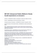
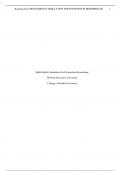
Actin monomer assemblies
Actin exists as a Globular monomer called G-actin. These monomers linearly assemble reversibly to
form F-actin: a filamentous actin polymer.
• each actin molecule contains a Mg2+ ion complexed with either ATP or ADP → actin is an
ATPase → only assembles when ATP-bound
G-actin denatures very quickly when not ATP/ADP bound or Mg2+ bound
• F-actin made of two helices of actin subunits
• F-actin has structural and functional polarity
− all subunits are oriented the same way → filament exhibits polarity: different ends
one end favours subunit addition (designated +)
other favours subunit dissociation (-)
→ ATP-binding clefts contact neighbouring subunits, except for – end
F-actin identification among cytoskeletal fibres by arrowhead decoration → myosin S1 binds to actin
only
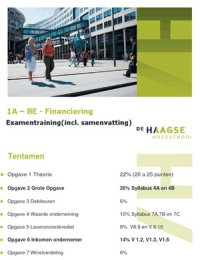
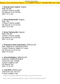
Dynamics of actin filaments
Structure oa regulated by influencing the stability causing spatially and temporarily controlled
polarisation and depolarisation. Pure G-actin polarisation proceeds in three sequential phases:
1. Nucleation phase: lag period in which subunits combine into oligomers of 2-3 subunits →
template for 2.
2. Elongation phase: oligomer length increase by both end addition → G-actin conc. decrease
until steady state reached: See physical organic chem 1 for polymerisation kinetics
3. steady-state phase: G-actin exchange on both ends but no net change in total length
The critical concentration Cc is the concentration at which filaments are formed → at steady state,
the conc. of monomeric actin remains at Cc
• subunit addition to both ends is asymmetric → (+) end grows 10 times faster than (-) end
• dissociation rate both ends nearly equal
Cc+ is the critical concentration for the (+) end: at this point the rate of assembly is equal to the rate
of disassembly at (+)
• If C > Cc+ net growth at (+) end and vise versa
rate of disassembly is independent of the free ATP-G-actin concentration
Cc- is the critical conc. for the (-) end: rate of assembly at (-) = rate of disassembly (-)
• if C > Cc- net growth at (-) and vise versa
• rate of disassembly almost equal to assembly naturally
Cc+ < Cc- because association at the (+) occurs much faster, so a lower conc. is necessary to equalise
dissociation with association at this point.
• if C = Cc-, C > Cc+ so if Cc+ < C < Cc-, steady state is reached because the (+) grows whereas
the (-) disassembles
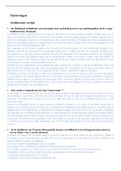
→ this is called treadmilling
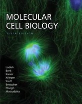
Treadmilling is powered by hydrolysis of ATP as only G-actin-ATP can assemble
, • Because of this process and treadmilling (+) has
G-ATP, followed by G-ADP-Pi, followed by G-ADP
at the (-) end
• G-ADP has undergone a conform. change by
disassembly of Pi that increases G-ADP
dissociation rate compared to G-ATP → this
underlies the faster dissociation at (-)
− in vivo, also ATP-G supply influences
kinetics
F-actin formation phases: nucleation, elongation, steady
state
The amounts of G-ATP/ADP+Pi/ADP in a filament are regulated as this determines the filament
shape.
Note that ATPase activity of actin and Pi release are slow, so if the actin assembly rate increases, the
amount of G-ATP at (+) increases too → therefore assembly is regulated by mechanisms
Faster treadmilling occurs in vivo then in vitro with pure minimally required
substances → treadmilling rate-limiting step for (-) end the rls for (+) end have
increased rate
• RLT (+) end is G-ADP into G-ATP conversion: catalysed by profilin →
binds the nucleotide-binding cleft so it catalyses ADP disassembly and
blocks binding at (-)
→ it also binds proteins rich in prolines during actin binding
− new rate limiting step is now the AVAILABILITY of ATP-G, such
that controlling this controls the growth rate (+)
o thymosin-β4 reversibly binds G-ATP, thereby inhibiting
its association
• RLT (-) end is G-ATP hydrolysis: catalysed by cofilin → binds specifically
F-actin ADP and destabilises twists bridging two actin subunits
Capping proteins block assembly and disassembly at actin filament (+) and (-) to spatially and timely
regulate filament growth
• The (+) side binds a protein known as CapZ → generally sufficient CapZ conc. to rapidly cap
newly formed (+) ends: so CapZ activity needs to be regulated by two mechanisms to allow
filament growth/treadmilling
− Regulatory phospholipid
phosphatidylinositol 4,5-
bisphosphate PIP2
− Regulatory proteins that bind (+)
to avoid CapZ binding but allow
G-ATP assembly
• The (-) side binds tropomodulin that
inhibits mostly disassembly → found
predominantly in cells that need highly
stabilised actin filaments
− binds tropomyosin that lies along the filament as well as actin
,Chapter 1 Cytoskeleton: microfilaments
This chapter surrounds the question how diversity of cellular shape, organisation and motility is
achieved.
The cytoskeleton
Cells must have different protein combinations in order to carry out their specific functions. Often
cells have functionally distinct regions, a phenomenon that the cell achieves by cell polarity.
• the cell’s functional polarity and internal organisation are provided by the cytoskeleton: a 3D
filamentous protein network
− the cytoskeleton is attached to the plasma membrane and internal organelles →
framework for cellular organisation
− the cytoskeleton is very dynamic → reorganisation: lengths and dynamics + local
regulation
The cytoskeleton provides support and organisation. It is composed of three major filament systems.
each system is a polymer of assembled subunits → subunits regulated in time and space.
• Microfilaments: actin polymers → organised by actin binding proteins
− important in plasma membrane organisation: shape surface structures
− MFs can serve on their own or be tracks for ATP-powered myosin motor proteins →
contraction/cargo transport
• Microtubules: tubulin polymers → organised by MT-associated proteins
− provide organisational framework for cell organelles and structural support
− mitotic spindle formation
− tracks for ATP-driven cargo transport via kinesins
• Intermediate filaments: tissue-specific filamentous structures → serve many different
functions
− barrier functions, nuclear structure support
− not used as tracks by motor proteins
Cytoskeletal arrangements very between cell types → accomplished by cellular signalling for
organisation: structure determines function
• signalling may be from the ECM → signal transduction via receptors
Microfilaments and actin structures
Due to the structure-function relationship and regulated structure, microfilaments have lots of
structures that all underly particular cellular functions (via actin)
• structural role
• harnessing power of actin polymerisation to do work
• tracks for myosin motors
Actin isoforms
Actin-binding proteins arrange MF by assembling actin. Microfilaments: actin polymers + associated
proteins.
• Humans have six actin genes: each encodes a different isoform → three groups of isoforms,
each with a different functions, therefore abundant in specific regions of the cell or in
proteins with specific functions
, • α-actins: contractile
• β-actins: cell cortex and leading edge of motile cells
• γ-actins: stress fibers
Actin monomer assemblies
Actin exists as a Globular monomer called G-actin. These monomers linearly assemble reversibly to
form F-actin: a filamentous actin polymer.
• each actin molecule contains a Mg2+ ion complexed with either ATP or ADP → actin is an
ATPase → only assembles when ATP-bound
G-actin denatures very quickly when not ATP/ADP bound or Mg2+ bound
• F-actin made of two helices of actin subunits
• F-actin has structural and functional polarity
− all subunits are oriented the same way → filament exhibits polarity: different ends
one end favours subunit addition (designated +)
other favours subunit dissociation (-)
→ ATP-binding clefts contact neighbouring subunits, except for – end
F-actin identification among cytoskeletal fibres by arrowhead decoration → myosin S1 binds to actin
only
Dynamics of actin filaments
Structure oa regulated by influencing the stability causing spatially and temporarily controlled
polarisation and depolarisation. Pure G-actin polarisation proceeds in three sequential phases:
1. Nucleation phase: lag period in which subunits combine into oligomers of 2-3 subunits →
template for 2.
2. Elongation phase: oligomer length increase by both end addition → G-actin conc. decrease
until steady state reached: See physical organic chem 1 for polymerisation kinetics
3. steady-state phase: G-actin exchange on both ends but no net change in total length
The critical concentration Cc is the concentration at which filaments are formed → at steady state,
the conc. of monomeric actin remains at Cc
• subunit addition to both ends is asymmetric → (+) end grows 10 times faster than (-) end
• dissociation rate both ends nearly equal
Cc+ is the critical concentration for the (+) end: at this point the rate of assembly is equal to the rate
of disassembly at (+)
• If C > Cc+ net growth at (+) end and vise versa
rate of disassembly is independent of the free ATP-G-actin concentration
Cc- is the critical conc. for the (-) end: rate of assembly at (-) = rate of disassembly (-)
• if C > Cc- net growth at (-) and vise versa
• rate of disassembly almost equal to assembly naturally
Cc+ < Cc- because association at the (+) occurs much faster, so a lower conc. is necessary to equalise
dissociation with association at this point.
• if C = Cc-, C > Cc+ so if Cc+ < C < Cc-, steady state is reached because the (+) grows whereas
the (-) disassembles
→ this is called treadmilling
Treadmilling is powered by hydrolysis of ATP as only G-actin-ATP can assemble
, • Because of this process and treadmilling (+) has
G-ATP, followed by G-ADP-Pi, followed by G-ADP
at the (-) end
• G-ADP has undergone a conform. change by
disassembly of Pi that increases G-ADP
dissociation rate compared to G-ATP → this
underlies the faster dissociation at (-)
− in vivo, also ATP-G supply influences
kinetics
F-actin formation phases: nucleation, elongation, steady
state
The amounts of G-ATP/ADP+Pi/ADP in a filament are regulated as this determines the filament
shape.
Note that ATPase activity of actin and Pi release are slow, so if the actin assembly rate increases, the
amount of G-ATP at (+) increases too → therefore assembly is regulated by mechanisms
Faster treadmilling occurs in vivo then in vitro with pure minimally required
substances → treadmilling rate-limiting step for (-) end the rls for (+) end have
increased rate
• RLT (+) end is G-ADP into G-ATP conversion: catalysed by profilin →
binds the nucleotide-binding cleft so it catalyses ADP disassembly and
blocks binding at (-)
→ it also binds proteins rich in prolines during actin binding
− new rate limiting step is now the AVAILABILITY of ATP-G, such
that controlling this controls the growth rate (+)
o thymosin-β4 reversibly binds G-ATP, thereby inhibiting
its association
• RLT (-) end is G-ATP hydrolysis: catalysed by cofilin → binds specifically
F-actin ADP and destabilises twists bridging two actin subunits
Capping proteins block assembly and disassembly at actin filament (+) and (-) to spatially and timely
regulate filament growth
• The (+) side binds a protein known as CapZ → generally sufficient CapZ conc. to rapidly cap
newly formed (+) ends: so CapZ activity needs to be regulated by two mechanisms to allow
filament growth/treadmilling
− Regulatory phospholipid
phosphatidylinositol 4,5-
bisphosphate PIP2
− Regulatory proteins that bind (+)
to avoid CapZ binding but allow
G-ATP assembly
• The (-) side binds tropomodulin that
inhibits mostly disassembly → found
predominantly in cells that need highly
stabilised actin filaments
− binds tropomyosin that lies along the filament as well as actin