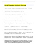
ECG interpretation
SA NODE 1
BACHMANN 'S
INTERMODAL
'
- BUNDLE
÷.
*.
¥÷÷?
Avnoae
AV NODE LEFT BUNDLE
brancnµ ÷
Bundle of His -
BRANCH
* "
BUNDLE OF HIS
RIGHT BUNDLE
BRANCH PURKINJE FIBERS
QRS
complex
I t
R
P wave
-
> 0 . 08 -
0.10 sec
PR interval -
> 0.12 -
0.20 sec ( 3- 5 smsq )
QRS complex
-
> 0 . 08 -
0.120sec ( 2- 3 sm sq )
P PR ST T QT interval -
> 0.35 -
0.43 sec
geameint , Segni U
ST segment
-
> 0.080 -
0.120sec ( 80 -
120msec )
T wave
-
> 0.10 -
0.25 sec +
" "
1
PR interval
Q 1ST interval U > =3 T same lead
wave
usually
-
wave amp in
S
QT interval
-
# 5mm aooms
1-
Bias r
→ -
>
(
CBB )
④
"
ate :# of P 10
waves 6
)
in sec ✗
1sec ( best for
↳ 5 BB
irregular)
=
)
(
or : 300
No BB btwn two R 's ( best for
regular )
> 1mm 40ms
( )
-
>
-
-
, ,
, The 12 Views of the Heart
• Standard EKG consists of 12 leads placed on body.
• Each lead “views” the heart from different angle.
• Two electrodes on arms and 2 on legs; these serve 6 limb leads (3 standard and 3 augmented).
• Six electrodes on chest for 6 precordial leads.
• Standard positioning of electrodes allows comparison between EKGs.
THE SiX LiMB LEADS
• View heart in frontal (vertical) plane.
• Each electrode is designated positive or negative.
• Each lead has unique angle of orientation to view heart
THE SiX PRECORDiAL LEADS
• View heart in horizontal plane.
• Record forces moving anteriorly and posteriorly.
• Each of six leads is made positive in turn; body is ground.
• V1 directly over right ventricle (right ventricular)
• V2, V3 over interventricular septum (anterior)
• V4 over apex of left ventricle (anterior)
• V5, V6 over lateral left ventricle (left lateral
÷ ÷ ¥ ¥E¥ ¥ ÷