Large vessel vasculitis
Introduction
Large arteries are the aorta and its major branches, medium arteries defined as the main
visceral arteries and their branches, small vessels defined as small intraparenchymal
arteries, arterioles, capillaries and venules.
Anatomy of large vessels: intima (endothelial cells), media (smooth muscle cells + connective
tissue), adventitia (vasa vasorum). The adventitia houses the vasa vasorum, DCs, fibroblasts
and nerve endings.
In contrast to the necrotizing vasculitides affecting the small- and medium-sized vessels, the
large-vessel vasculitides are:
-not necrotizing
-not dominated by neutrophil infiltration, but by monocytes, macrophages, T cells
-granulomatous (also GPA, unlike Behçet).
GCA
Overall GCA is the most common systemic vasculitis, with a lifetime risk of up to 1% in USA
and incidence 1-27/100.000 individuals ≥50yrs. Granulomatous, non-necrotizing, large (and
medium) vessels. Exclusively in >50yrs, women 2-3x > men. Smoking increases the risk for
GCA 6x in women. Having diabetes reduces the risk of GCA by 50% in women. Caucasians
(Scandinavian origin) > Black, Hispanic. 60% of GCA patients have HLA-DRB1*04. GCA is the
form of systemic vasculitis most closely associated with HLA-II genes. Susceptibility to GCA
and PMR has also been associated with polymorphisms of genes for TNF, ICAM and IL-18.
Anatomy
Temporal artery arises from the external carotid (on the level of the parotid), passes
anterior to the ear and splits into frontal and parietal branch.
Aorta: 3 branches: i) right brachionocephalic (splits into right common carotid + right
subclavian), ii) left common carotid, iii) left subclavian.
Vertebral arteries stem from the subclavian arteries.
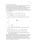
Criteria
,Note that MRI is not in the criteria, neither are LV symptoms (they refer to TAK), only aorta in
PET.
Histology
Biopsy: at least 1cm of vessel length. We perform temporal artery biopsy within 2 wks (max.
4 wks) of treatment, ideally within 1 week.
False-negative biopsy
-skip lesions
-focal, circumscribed lesion (inflammatory changes may be missed if only one
section is examined, so =we examine at least 3 slides)
-treatment (GC reduce the sensitivity)
-chronicity
-in cases of only LV involvement (only 50% will be positive)
Histologic findings
1) Inflammation can be limited to the adventitia, extending into the media,
transmural or affecting the adventitia and intima sparing the media (‘concentric rings’).
Infiltrate is dominated by CD4+ and macrophages.
2) Intimal thickening – lumen occlusion
3) Granulomas within the wall (histiocytes and multinucleated giant cells) (in 25-
50%) (no granuloma formation in tissue, like GPA, sarcoidosis)
4) Destruction of vessel wall layers (esp. internal elastic lamina)
5) In some patients (15%), the inflammation is restricted to the periadventitial small
vessels or to the adventitial vasa vasorum.
, As the medial smooth muscle cell layer loses thickness, the intima becomes hyperplastic,
compromising or occluding the arterial lumen. Although the vessel lumen may become
critically narrowed, thrombosis is not the central event. Hyperplasia and scarring are
irreversible. The “classic” pattern (only seen in 50%) includes granulomatous inflammation
of the inner half of the media, centered on the internal elastic lamina, with a mononuclear
infiltrate, multinucleate giant cells and fragmentation of the internal elastic lamina. Giant
cells occur in only 50%. In the other 50%, granulomas and giant cells are absent and a
nonspecific panarteritis is seen, including mixed inflammatory infiltrates composed largely of
CD4+ and macrophages mixed with a few eosinophils; neutrophils are rare. Fibrinoid necrosis
should not be seen (GCA is NOT necrotizing) and if present, should suggest an alternate
diagnosis (ex. GPA).
Sometimes biopsy shows structural degenerative changes in the absence of
inflammation, such as media-intimal scar, intimal hyperplasia, fragmentation of internal
elastic lamina, calcifications or fibrosis of the adventitia. However, these changes are not
specific of GCA because they are also found in healthy older adults.
Pathophysiology
Because GCA affects vessels with an internal elastic lamina and vasa vasorum and because
intracranial arteries lose these structures after penetrating the dura, it is not surprising that
GCA rarely involves intracranial arteries.
The arterial wall is an immunoprivileged site. This immunoprivilege is lost with age
as older adults demonstrate adventitial lymphoid infiltrates, sometimes aggregated in
lymphoid follicles (‘artery tertiary lymphoid organs’).
The major vascular abnormality leading to clinical disease is a non-thrombotic
luminal occlusion, caused by rapid and concentric growth of the intima. Intimal hyperplasia
is generated by the mobilization of smooth muscle cells, their directed migration towards
the lumen and their proliferation and matrix deposition.
T-cell–mediated immunopathology
DCs, CD4+ (Th1, Th17) and macrophages. B cells are not found within the arterial wall; no
pathognomonic antibodies have been identified and hypergammaglobulinemia is absent.
However, changes of B-cells in the blood and B-cell infiltrates including ectopic lymphoid
structures have been found in the temporal arteries and aortic tissue of patients with GCA,
implying a role for B cells in the effector phase of GCA.
Adventitia is the only layer that can be penetrated by vasa vasorum and DCs lay there.
1) Immature DCs, lacking the costimulatory molecules CD80/86, are normally
present in the adventitia of arteries and because of a lack of costimulation, tolerate T-cells
that recognize (auto)antigens presented by these DCs. PAMPs/DAMPs may activate the DCs
and they release chemokines that recruit T-cells and macrophages into the vessel wall and
also produce cytokines such as IL-6, IL-18, IL-23.
DCs in GCA are:
-primed (high expression of MHC-II, CD80/86 – binds to T-cell CD28)
-show an increased expression of TLR2 and TLR4
-PD-L1 deficient.
These contribute to T cell activation in vessel wall.
2) Recruited CD4+ interact with these mature DCs and are polarized into Th1 cells (in
the presence of IL-12, IL-18), Th17 cells (in the presence of IL-6, IL-1β, IL-23) or both, which
leads to a chronic inflammatory response.
-Th1 produce IFN-γ
-Th17 produce IL-17, IL-22
Decreased expression of PD-L1 on DCs in GCA prevents cessation of the CD4 response.
Introduction
Large arteries are the aorta and its major branches, medium arteries defined as the main
visceral arteries and their branches, small vessels defined as small intraparenchymal
arteries, arterioles, capillaries and venules.
Anatomy of large vessels: intima (endothelial cells), media (smooth muscle cells + connective
tissue), adventitia (vasa vasorum). The adventitia houses the vasa vasorum, DCs, fibroblasts
and nerve endings.
In contrast to the necrotizing vasculitides affecting the small- and medium-sized vessels, the
large-vessel vasculitides are:
-not necrotizing
-not dominated by neutrophil infiltration, but by monocytes, macrophages, T cells
-granulomatous (also GPA, unlike Behçet).
GCA
Overall GCA is the most common systemic vasculitis, with a lifetime risk of up to 1% in USA
and incidence 1-27/100.000 individuals ≥50yrs. Granulomatous, non-necrotizing, large (and
medium) vessels. Exclusively in >50yrs, women 2-3x > men. Smoking increases the risk for
GCA 6x in women. Having diabetes reduces the risk of GCA by 50% in women. Caucasians
(Scandinavian origin) > Black, Hispanic. 60% of GCA patients have HLA-DRB1*04. GCA is the
form of systemic vasculitis most closely associated with HLA-II genes. Susceptibility to GCA
and PMR has also been associated with polymorphisms of genes for TNF, ICAM and IL-18.
Anatomy
Temporal artery arises from the external carotid (on the level of the parotid), passes
anterior to the ear and splits into frontal and parietal branch.
Aorta: 3 branches: i) right brachionocephalic (splits into right common carotid + right
subclavian), ii) left common carotid, iii) left subclavian.
Vertebral arteries stem from the subclavian arteries.
Criteria
,Note that MRI is not in the criteria, neither are LV symptoms (they refer to TAK), only aorta in
PET.
Histology
Biopsy: at least 1cm of vessel length. We perform temporal artery biopsy within 2 wks (max.
4 wks) of treatment, ideally within 1 week.
False-negative biopsy
-skip lesions
-focal, circumscribed lesion (inflammatory changes may be missed if only one
section is examined, so =we examine at least 3 slides)
-treatment (GC reduce the sensitivity)
-chronicity
-in cases of only LV involvement (only 50% will be positive)
Histologic findings
1) Inflammation can be limited to the adventitia, extending into the media,
transmural or affecting the adventitia and intima sparing the media (‘concentric rings’).
Infiltrate is dominated by CD4+ and macrophages.
2) Intimal thickening – lumen occlusion
3) Granulomas within the wall (histiocytes and multinucleated giant cells) (in 25-
50%) (no granuloma formation in tissue, like GPA, sarcoidosis)
4) Destruction of vessel wall layers (esp. internal elastic lamina)
5) In some patients (15%), the inflammation is restricted to the periadventitial small
vessels or to the adventitial vasa vasorum.
, As the medial smooth muscle cell layer loses thickness, the intima becomes hyperplastic,
compromising or occluding the arterial lumen. Although the vessel lumen may become
critically narrowed, thrombosis is not the central event. Hyperplasia and scarring are
irreversible. The “classic” pattern (only seen in 50%) includes granulomatous inflammation
of the inner half of the media, centered on the internal elastic lamina, with a mononuclear
infiltrate, multinucleate giant cells and fragmentation of the internal elastic lamina. Giant
cells occur in only 50%. In the other 50%, granulomas and giant cells are absent and a
nonspecific panarteritis is seen, including mixed inflammatory infiltrates composed largely of
CD4+ and macrophages mixed with a few eosinophils; neutrophils are rare. Fibrinoid necrosis
should not be seen (GCA is NOT necrotizing) and if present, should suggest an alternate
diagnosis (ex. GPA).
Sometimes biopsy shows structural degenerative changes in the absence of
inflammation, such as media-intimal scar, intimal hyperplasia, fragmentation of internal
elastic lamina, calcifications or fibrosis of the adventitia. However, these changes are not
specific of GCA because they are also found in healthy older adults.
Pathophysiology
Because GCA affects vessels with an internal elastic lamina and vasa vasorum and because
intracranial arteries lose these structures after penetrating the dura, it is not surprising that
GCA rarely involves intracranial arteries.
The arterial wall is an immunoprivileged site. This immunoprivilege is lost with age
as older adults demonstrate adventitial lymphoid infiltrates, sometimes aggregated in
lymphoid follicles (‘artery tertiary lymphoid organs’).
The major vascular abnormality leading to clinical disease is a non-thrombotic
luminal occlusion, caused by rapid and concentric growth of the intima. Intimal hyperplasia
is generated by the mobilization of smooth muscle cells, their directed migration towards
the lumen and their proliferation and matrix deposition.
T-cell–mediated immunopathology
DCs, CD4+ (Th1, Th17) and macrophages. B cells are not found within the arterial wall; no
pathognomonic antibodies have been identified and hypergammaglobulinemia is absent.
However, changes of B-cells in the blood and B-cell infiltrates including ectopic lymphoid
structures have been found in the temporal arteries and aortic tissue of patients with GCA,
implying a role for B cells in the effector phase of GCA.
Adventitia is the only layer that can be penetrated by vasa vasorum and DCs lay there.
1) Immature DCs, lacking the costimulatory molecules CD80/86, are normally
present in the adventitia of arteries and because of a lack of costimulation, tolerate T-cells
that recognize (auto)antigens presented by these DCs. PAMPs/DAMPs may activate the DCs
and they release chemokines that recruit T-cells and macrophages into the vessel wall and
also produce cytokines such as IL-6, IL-18, IL-23.
DCs in GCA are:
-primed (high expression of MHC-II, CD80/86 – binds to T-cell CD28)
-show an increased expression of TLR2 and TLR4
-PD-L1 deficient.
These contribute to T cell activation in vessel wall.
2) Recruited CD4+ interact with these mature DCs and are polarized into Th1 cells (in
the presence of IL-12, IL-18), Th17 cells (in the presence of IL-6, IL-1β, IL-23) or both, which
leads to a chronic inflammatory response.
-Th1 produce IFN-γ
-Th17 produce IL-17, IL-22
Decreased expression of PD-L1 on DCs in GCA prevents cessation of the CD4 response.