BLOK 1.4
Case elaborations
Thinking and doing
, Case 1
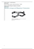
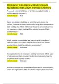
Evolution of the brain
Gee Die Messen 172et 172ij Te
Di
Front brain Me
· ·
Mid brain
M
Hind brain
= Spinal cord M
Sp
3 week old brain 6 week old brain
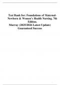
Brain lobes
The brain is made up of 2 halves (hemispheres), which can be divided in lobes:
• frontal lobe (lobus frontalis) → coordinates cognitive, emotional and motivational processes such as planning and
impulse control.
• parietal lobe (lobus parietalis) → coordinates senses and cognitive functions, such as attention, special awareness,
reading and calculating.
• temporal lobe (lobus temporalis) → consists of the additive cortex and works close together with the hippocampus
(front of the lobe), it also contains the centre of wernicke.
• occipital lobe (lobus occipitalis) → coordinates sight
The brain has multiple folds, these are called gyri. There are a couple of sulci that are
The are separated by 2 types of grooves; • central sulcus → separates the
• sulci → un-deep grooves • lateral sulcus → separates the
• fissure → deep grooves • parieto-occipital sulcus → sepa
Brain cortex
, Small brain
The small brain, also known as the cerebellum, lies behind the pons and right under the
occipital lobe. it consists of 2 parts, divided by a medial line (the vermis). The activity of the
cerebellum isn't regulated by will. The cerebellum regulates the muscles movement and the
balance. The deep nucleii of the cerebellum are:
1. Nucleus dentatus → fine coordination of movement
2. Nucleus emboliformis and nucleus globosus → muscle tone and rough movements
3. Nucleus fastigii → regulates balance and posture
Basal ganglia
The basal ganglia is a ring-form structure around the thalamus, together with the cerebellum they form a
control loop to regulate the motor activity which generates from the cortex. The signal from the cortex gets
inhibited, excreted or managed to make certain movements easier. it consists of the following things:
• nucleus caudatus
• putamen
• globus pallidus (pale nucleus)
• nucleus subtalamicus
• nucleus ruber
Ventricle system
The brain ventricles are connected with the central canal of the spinal cord, canalis centralis. The hollow
ventricular spaces are filled with cerebrospinal fluid (liquor cerebrospinalis) and lined with ependymcells, a
type of neurogliacells. Some lateral ventricles lie close to each other so they get separated by the septum
pellucidum, a thin membrane.
Every lateral ventricle communicates with a small third ventricle (ventriculus tertius), this is laid in the
diencephalon, through a channel called the foramen intraventriculairis (foramen of Monro). The third
ventricle is connected to the fourth ventricle (ventriculus quartus) via a channel called the cerebral
aquaduct, which leads to the midbrain. This is connected to the central channel of the spinal cord. There are
3 holes marking the walls of the 4th ventricle; 2 lateral apertures on the sides of the ventricles and the
median aperture on the top of the ventricle. These apertures connect the ventricles to the subarachnoidal
space, a with fluid filled space around the brain. From this space the liquor travels to the arachnoidal villi
(pacchioni granules ) which leads to the big venous sinus (sinus saggital superior), the excess fluid gets
absorbed by the venous system
Liquor cerebrospinalis gets produced by ependym-cells who lie in the walls of the ventricles and in the plexus choroideus of
any type of shock from blunt force trauma and it drains waist products
Areas of brodmann
The areas of brodmann are areas in the brain cortex, it consists of 52 different areas but only a few are important:
Case elaborations
Thinking and doing
, Case 1
Evolution of the brain
Gee Die Messen 172et 172ij Te
Di
Front brain Me
· ·
Mid brain
M
Hind brain
= Spinal cord M
Sp
3 week old brain 6 week old brain
Brain lobes
The brain is made up of 2 halves (hemispheres), which can be divided in lobes:
• frontal lobe (lobus frontalis) → coordinates cognitive, emotional and motivational processes such as planning and
impulse control.
• parietal lobe (lobus parietalis) → coordinates senses and cognitive functions, such as attention, special awareness,
reading and calculating.
• temporal lobe (lobus temporalis) → consists of the additive cortex and works close together with the hippocampus
(front of the lobe), it also contains the centre of wernicke.
• occipital lobe (lobus occipitalis) → coordinates sight
The brain has multiple folds, these are called gyri. There are a couple of sulci that are
The are separated by 2 types of grooves; • central sulcus → separates the
• sulci → un-deep grooves • lateral sulcus → separates the
• fissure → deep grooves • parieto-occipital sulcus → sepa
Brain cortex
, Small brain
The small brain, also known as the cerebellum, lies behind the pons and right under the
occipital lobe. it consists of 2 parts, divided by a medial line (the vermis). The activity of the
cerebellum isn't regulated by will. The cerebellum regulates the muscles movement and the
balance. The deep nucleii of the cerebellum are:
1. Nucleus dentatus → fine coordination of movement
2. Nucleus emboliformis and nucleus globosus → muscle tone and rough movements
3. Nucleus fastigii → regulates balance and posture
Basal ganglia
The basal ganglia is a ring-form structure around the thalamus, together with the cerebellum they form a
control loop to regulate the motor activity which generates from the cortex. The signal from the cortex gets
inhibited, excreted or managed to make certain movements easier. it consists of the following things:
• nucleus caudatus
• putamen
• globus pallidus (pale nucleus)
• nucleus subtalamicus
• nucleus ruber
Ventricle system
The brain ventricles are connected with the central canal of the spinal cord, canalis centralis. The hollow
ventricular spaces are filled with cerebrospinal fluid (liquor cerebrospinalis) and lined with ependymcells, a
type of neurogliacells. Some lateral ventricles lie close to each other so they get separated by the septum
pellucidum, a thin membrane.
Every lateral ventricle communicates with a small third ventricle (ventriculus tertius), this is laid in the
diencephalon, through a channel called the foramen intraventriculairis (foramen of Monro). The third
ventricle is connected to the fourth ventricle (ventriculus quartus) via a channel called the cerebral
aquaduct, which leads to the midbrain. This is connected to the central channel of the spinal cord. There are
3 holes marking the walls of the 4th ventricle; 2 lateral apertures on the sides of the ventricles and the
median aperture on the top of the ventricle. These apertures connect the ventricles to the subarachnoidal
space, a with fluid filled space around the brain. From this space the liquor travels to the arachnoidal villi
(pacchioni granules ) which leads to the big venous sinus (sinus saggital superior), the excess fluid gets
absorbed by the venous system
Liquor cerebrospinalis gets produced by ependym-cells who lie in the walls of the ventricles and in the plexus choroideus of
any type of shock from blunt force trauma and it drains waist products
Areas of brodmann
The areas of brodmann are areas in the brain cortex, it consists of 52 different areas but only a few are important: