Echocardiogram
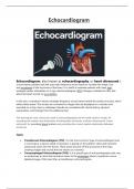
Echocardiogram, also known as echocardiography, or heart ultrasound is
a noninvasive, painless test that uses high-frequency sound waves to visualize the shape, size,
and movement of the structures of the heart. It is useful to evaluate patients with chest pain,
enlarged cardiac silhouettes on X-rays, electrocardiogram (ECG) changes unrelated to CAD, and
abnormal heart sounds on auscultation.
In this test, a transducer directs ultrahigh-frequency sound waves toward the cardiac structure, which
reflect these waves. The echoes are converted to images that are displayed on a monitor and
recorded on a strip chart or videotape. Results are correlated with clinical history, physical
examination, and findings from the additional test.
The techniques most commonly used in echocardiography are M-mode (motion mode), for
recording the motion and dimensions of intracardiac structures, and two-dimensional (cross-
sectional), for recording lateral motion and providing the correct spatial relationship between
structures.
Types
Transthoracic Echocardiogram (TTE). It is the most common type of echocardiogram and
is noninvasive. A device called a transducer is placed on the patient’s chest and transmits
ultrasound waves into the thorax. These waves bounce off the structures of the heart,
creating images and sounds that are shown on a monitor.
Transesophageal Echocardiogram (TOE). It is a special type of echocardiography that uses
an endoscope to assist the transducer down to the esophagus where it produces a more
detailed image of the heart than a transthoracic echocardiogram.
, Stress Echocardiogram. An echocardiogram is performed while the patient is using a
treadmill or stationary bicycle. This type can be used to measure the function of the heart
both at rest and while exercising.
Dobutamine Stress Echocardiogram. For patients who are unable to exercise on a
treadmill, a drug called dobutamine is given instead through a vein that stimulates the heart
in a similar manner as exercise. This type of echocardiogram is used to evaluate coronary
artery disease and measures the effectiveness of a cardiac therapeutic regimen.
Doppler echocardiogram. Measures and assess the blood flow through the heart and blood
vessels.
Indication
Detect and evaluate valvular abnormalities
Detect atrial tumors
Measure the size of the heart chambers
Evaluate chambers and valves in congenital heart disorders
Diagnose hypertrophic and related cardiomyopathies
Evaluate cardiac function or wall motion after myocardial infarctions
Detect pericardial effusion and mural thrombi
Procedure
The following are the steps and processes of how an echocardiography or echocardiogram is
performed:
1. Place the patient in a supine position.
Patient is placed in a supine position and a conductive gel is applied to the third or fourth
intercostal space to the left of the sternum. The transducer is placed directly over it.
2. Transducer is placed
The transducer directs ultra-high-frequency sound waves towards cardiac structures, which
reflect these waves; the transducer picks up the echoes, converts them to electrical impulses,
and relays them to an echocardiography machine for display.
3. Motion mode is used
In motion mode (M-mode), a single, pencil-like ultrasound beam strikes the heart and
produces a vertical view, which is useful for recording the motion and dimensions of
intracardiac structures.
4. Change in position
In two-dimensional echocardiography, a cross-sectional view of the cardiac structures is used
for recording the lateral motion and spatial relationship between structures. For a left lateral
view, the patient is placed on his left side.