BMET5907- Semester 2 Notes:
L1: Course overview, anatomy review, bone and joints.
Skeletal System:
The main role of the skeletal system is to provide:
1. Structure and stability for muscle
2. Protection for soft tissue (e.g. spinal cord and liver)
3. Storage of calcium and phosphate ions to improve hardness and strength
4. Blood cell production
*BONES NEVER LIE ® remodels according to environmental stimulus
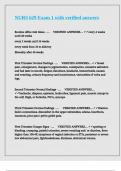
Components of the Skeletal System (Axial + Appendicular)
Axial Bones Function
Skull (22) Cranial vault of 8 bones + 14 • Protect brain
facial bones • Supports facial muscle and muscle of mastication
• Support airways and swallowing muscle
• Auditory ossicles in inner ear amplify sound for hearing
Vertebrae • Cervical (C1-7) • Protects spinal cord
• Thoracic (T1-12) • Bones gets less dense when moving down from head
• Lumbar (L1-5) *Note: very complex and varies between patient
• Sacral (S1-5)
• Coccyx (4)
Ribs (12) • 7 ribs join sternum • Enclose lungs and heart
directly • Protect upper abdominal structures (liver, spleen, pancreas)
• 3 false ribs (join to 7th) • Attachment for diaphragm + act to expand lungs by swinging up
• 2 floaters like a bucket handle
Appendicular Bones Function
Shoulder Girdle Scapula and Girdles Attach and pivot arms to axial skeleton (high deg.
Of freedom)
Arms • Humerus, radius/ulna,
• 8 carpal (wrist) bones
• 19 hand bones
Pelvic Girdle • 2 wings (each of 3 dfused bones)
• Houses sacrum
Legs • Femur, Tibia/fibula
• 7 Tarsals (ankle + midfoot) bones
• 19 foot bones
,Bone Structure and Shape
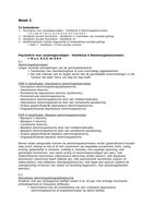
2 major types of bone structure
Type Properties Function
1. Cortical • Dense and strong • Usually located in shaft of long bones
• Low porosity (<30%) • Long distance weight and force
transmission in body
2. Cancellous • Lighter (1/3 density of cortical) • Bear weight (at ends of long bone) +
(spongy • 10x less stiffer (i.e. less strong per unit area) than aided by denser plates or trabeculae
bone) cortical BUT 5x more ductile within structure
• Less aligned in its structure • Alignment of plates enables better
• Higher porosity (30-90%) handling of loads from different
directions ® plates arranged in
• NO haversian canal
response to load dimension and
• Red marrow between trabecular plates = provides direction (Wolf’s law)
hydraulic stiffness and shock resistance
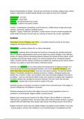
4 major types of bone shape
Description Example
1. Long • Long and wide • Femur, tibia, humerus,
bones • Long cortical shaft (narrowest part of bone) ulna
• Broadening at each end (composed of cancellous bone) ® • Small long bones (e.g.
allows weight to be transferred from one bone to another via phalanges in fingers)
joints with a lower force/unit area compared to if smaller
diameter of cortical section was maintained
• Medullary cavity of long bones = marrow
2. Short • NO shaft • Talus (contained all
Bones • Composed of mostly cancellous bone + outer shell of cortical carpal bones)
bone • Patella (knee cap) ®
exists within a tendon
3. Flat • Thin layer of cortical bone either side of thin layer of cancellous • Skull
Bones bone • Sternum
• Act as Stiff plates able to handle and response to large loads • Scapula
applied by muscles attached to bones (while being light and
delicate)
4. Irregular • Cancellous bone surrounded by cortical bone • Vertebrae
bones • ossicles in inner ear
Parts of Bone Location
Diaphysis Long shaft of bone
Epiphysis Ends of bone
Metaphysis Between epiphysis and diaphysis (links 2 areas)
, Joints:
Mobility Description
Synarthroses • ALMOST NO • 2 bones held together by fibrous
relative motion tissue or cartilage
• E.g. skull bones ® which do bend
and flex to some extent during
chewing)
Amphiarthoses • Relatively little • 2 bones linked by fibrous disc or
motion (more ligaments
than • E.g. joints between vertebral bodies)
synarthroses)
Diarthroses • 2 bones freely • Joints have cavity + lined by synovial
movable tissue (filled with synovial fluid)
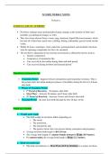
ANATOMICAL PLANES & DIRECTIONAL TERMS
Direction Opposite Direction
Anterior In front Posterior Back
Ventral (anterior) In front of spine Dorsal (posterior) Back of spine
Lateral Away from midline Medial Closer to midline
Distal Further from trunk Proximal Closer to Trunk (contextual and relative)
Superior Above Inferior Below
UNIQUE DIRECTIONS
Anterior-Posterior • Anterior-Posterior (AP) projection ® for unwell patients
vs. Posterior- who can’t stand
Anterior Projection
• Since heart is anterior structure ® magnified in AP view
(Why is labelling due to shorter distance between X-ray and patient
required)
• *Never consider the heart size to be enlarged if the
projection used is AP. If however the heart size is normal
on an AP view, then you can say it is not enlarged.
• Posterior-Anterior (PA) projection ®closer to real size
of heart
Medio-Lateral being a direction or axis from side to side or from median to
lateral.
L1: Course overview, anatomy review, bone and joints.
Skeletal System:
The main role of the skeletal system is to provide:
1. Structure and stability for muscle
2. Protection for soft tissue (e.g. spinal cord and liver)
3. Storage of calcium and phosphate ions to improve hardness and strength
4. Blood cell production
*BONES NEVER LIE ® remodels according to environmental stimulus
Components of the Skeletal System (Axial + Appendicular)
Axial Bones Function
Skull (22) Cranial vault of 8 bones + 14 • Protect brain
facial bones • Supports facial muscle and muscle of mastication
• Support airways and swallowing muscle
• Auditory ossicles in inner ear amplify sound for hearing
Vertebrae • Cervical (C1-7) • Protects spinal cord
• Thoracic (T1-12) • Bones gets less dense when moving down from head
• Lumbar (L1-5) *Note: very complex and varies between patient
• Sacral (S1-5)
• Coccyx (4)
Ribs (12) • 7 ribs join sternum • Enclose lungs and heart
directly • Protect upper abdominal structures (liver, spleen, pancreas)
• 3 false ribs (join to 7th) • Attachment for diaphragm + act to expand lungs by swinging up
• 2 floaters like a bucket handle
Appendicular Bones Function
Shoulder Girdle Scapula and Girdles Attach and pivot arms to axial skeleton (high deg.
Of freedom)
Arms • Humerus, radius/ulna,
• 8 carpal (wrist) bones
• 19 hand bones
Pelvic Girdle • 2 wings (each of 3 dfused bones)
• Houses sacrum
Legs • Femur, Tibia/fibula
• 7 Tarsals (ankle + midfoot) bones
• 19 foot bones
,Bone Structure and Shape
2 major types of bone structure
Type Properties Function
1. Cortical • Dense and strong • Usually located in shaft of long bones
• Low porosity (<30%) • Long distance weight and force
transmission in body
2. Cancellous • Lighter (1/3 density of cortical) • Bear weight (at ends of long bone) +
(spongy • 10x less stiffer (i.e. less strong per unit area) than aided by denser plates or trabeculae
bone) cortical BUT 5x more ductile within structure
• Less aligned in its structure • Alignment of plates enables better
• Higher porosity (30-90%) handling of loads from different
directions ® plates arranged in
• NO haversian canal
response to load dimension and
• Red marrow between trabecular plates = provides direction (Wolf’s law)
hydraulic stiffness and shock resistance
4 major types of bone shape
Description Example
1. Long • Long and wide • Femur, tibia, humerus,
bones • Long cortical shaft (narrowest part of bone) ulna
• Broadening at each end (composed of cancellous bone) ® • Small long bones (e.g.
allows weight to be transferred from one bone to another via phalanges in fingers)
joints with a lower force/unit area compared to if smaller
diameter of cortical section was maintained
• Medullary cavity of long bones = marrow
2. Short • NO shaft • Talus (contained all
Bones • Composed of mostly cancellous bone + outer shell of cortical carpal bones)
bone • Patella (knee cap) ®
exists within a tendon
3. Flat • Thin layer of cortical bone either side of thin layer of cancellous • Skull
Bones bone • Sternum
• Act as Stiff plates able to handle and response to large loads • Scapula
applied by muscles attached to bones (while being light and
delicate)
4. Irregular • Cancellous bone surrounded by cortical bone • Vertebrae
bones • ossicles in inner ear
Parts of Bone Location
Diaphysis Long shaft of bone
Epiphysis Ends of bone
Metaphysis Between epiphysis and diaphysis (links 2 areas)
, Joints:
Mobility Description
Synarthroses • ALMOST NO • 2 bones held together by fibrous
relative motion tissue or cartilage
• E.g. skull bones ® which do bend
and flex to some extent during
chewing)
Amphiarthoses • Relatively little • 2 bones linked by fibrous disc or
motion (more ligaments
than • E.g. joints between vertebral bodies)
synarthroses)
Diarthroses • 2 bones freely • Joints have cavity + lined by synovial
movable tissue (filled with synovial fluid)
ANATOMICAL PLANES & DIRECTIONAL TERMS
Direction Opposite Direction
Anterior In front Posterior Back
Ventral (anterior) In front of spine Dorsal (posterior) Back of spine
Lateral Away from midline Medial Closer to midline
Distal Further from trunk Proximal Closer to Trunk (contextual and relative)
Superior Above Inferior Below
UNIQUE DIRECTIONS
Anterior-Posterior • Anterior-Posterior (AP) projection ® for unwell patients
vs. Posterior- who can’t stand
Anterior Projection
• Since heart is anterior structure ® magnified in AP view
(Why is labelling due to shorter distance between X-ray and patient
required)
• *Never consider the heart size to be enlarged if the
projection used is AP. If however the heart size is normal
on an AP view, then you can say it is not enlarged.
• Posterior-Anterior (PA) projection ®closer to real size
of heart
Medio-Lateral being a direction or axis from side to side or from median to
lateral.