ECG interpretation
Demographics 1 small square = 0.04s = 40ms
1 big square = 0.2s = 200ms
Patient name, DOB, any symptoms (e.g. chest pain)
ECG date and time and which in series
Check calibration
o Paper speed – 25mm/s
o 1mV calibration deflection (at start of trace) – 2 large squares in height
Rate and rhythm
Use rhythm strip
Rate: 300 / number of large squares between R peaks OR, if irregular, total R waves on ECG x 6 (ECG is 10 seconds long)
o sinus bradycardia <60 (physical fitness; hypothermia; hypothyroidism; SA node disease; B-blockers)
o sinus tachycardia >100 (exercise/pain/anxiety; pregnancy; anaemia; PE; hypovolaemia; fever; thyrotoxicosis)
Rhythm
1. Regularity: mark 4 R peaks on plain piece of paper and move along trace to confirm (irregular may be: AF; ectopics; 2nd
degree AV block)
2. Sinus: look for a normal P wave before each QRS complex (no clear P waves and irregular QRS = AF; sawtooth baseline =
atrial flutter; broad complex tachycardia with no p waves = VF or VT or rarely SVT with BBB/WPW; narrow complex
tachycardia with abnormal or no p waves = supraventricular tachycardia)
Axis
Use leads I and II
Short method: QRS complexes in leads I and II are normally both predominantly positive
o If R waves point away from each other i.e. QRS predominantly positive in lead I and negative in lead II (‘legs apart’)
there is left axis deviation (i.e. more electricity going to left due to: LV hypertrophy/strain; left anterior hemiblock;
inferior MI; WPW; VT)
o If R waves point towards each other (‘legs together’ - right!) there is right axis deviation (i.e. more electricity going to
right due to: tall & thin body type; RV hypertrophy/strain e.g. in PE; left posterior hemiblock; lateral MI; WPW)
P wave
Use rhythm strip
Height ≤2 small squares (increased in right atrial hypertrophy e.g. caused by pulmonary hypertension or tricuspid stenosis)
Morphology
o Bifid = P mitrale (left atrial hypertrophy e.g. caused by mitral stenosis)
o Peaked = P pulmonale (right atrial hypertrophy)
PR interval
Use rhythm strip
Length 3-5 small squares
o Decreased: accessory conduction pathway
o Increased in AV block or ‘heartblock’
1st degree AV block: PR >5 small squares and regular
2nd degree AV block
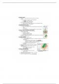
Mobitz type 1 (Wenkebach): PR progressively elongates until
there is failure of conduction of an atrial beat (then the cycle CARDIAC CONDUCTION CIRCUIT
1st and 2nd AV block may be repeats) SA node
caused by: ↑vagal tone/athletes,
coronary artery disease, Mobitz type 2: constant normal PR with occasional dropped
myocarditis, acute rheumatic beats
carditis, digoxin toxicity, or
electrolyte disturbances. 2nd degree AV block with 2:1/3:1/4:1 block: alternate AV node
Bundle of His LBB
conducted and non-conducted atrial beats (P:QRS) RBB
Anterior
3rd degree (complete) AV block: complete dissociation between p waves fascicle
and QRS complexes. Normal atrial beats which are not conducted to
ventricles resulting in ventricles self-depolarising at a much slower rate Posterior
‘ventricular escape rhythm’. fascicle
3rd degree AV block is caused by
fibrosis around Bundle of His
(may be caused by ischaemia,
congential, idiopathic, aortic
stenosis, or trauma) or block of
both bundle branches.
© 2014 Dr Christopher Mansbridge at www.OSCEstop.com, a source of free OSCE exam notes for medical students’ finals OSCE revision
Demographics 1 small square = 0.04s = 40ms
1 big square = 0.2s = 200ms
Patient name, DOB, any symptoms (e.g. chest pain)
ECG date and time and which in series
Check calibration
o Paper speed – 25mm/s
o 1mV calibration deflection (at start of trace) – 2 large squares in height
Rate and rhythm
Use rhythm strip
Rate: 300 / number of large squares between R peaks OR, if irregular, total R waves on ECG x 6 (ECG is 10 seconds long)
o sinus bradycardia <60 (physical fitness; hypothermia; hypothyroidism; SA node disease; B-blockers)
o sinus tachycardia >100 (exercise/pain/anxiety; pregnancy; anaemia; PE; hypovolaemia; fever; thyrotoxicosis)
Rhythm
1. Regularity: mark 4 R peaks on plain piece of paper and move along trace to confirm (irregular may be: AF; ectopics; 2nd
degree AV block)
2. Sinus: look for a normal P wave before each QRS complex (no clear P waves and irregular QRS = AF; sawtooth baseline =
atrial flutter; broad complex tachycardia with no p waves = VF or VT or rarely SVT with BBB/WPW; narrow complex
tachycardia with abnormal or no p waves = supraventricular tachycardia)
Axis
Use leads I and II
Short method: QRS complexes in leads I and II are normally both predominantly positive
o If R waves point away from each other i.e. QRS predominantly positive in lead I and negative in lead II (‘legs apart’)
there is left axis deviation (i.e. more electricity going to left due to: LV hypertrophy/strain; left anterior hemiblock;
inferior MI; WPW; VT)
o If R waves point towards each other (‘legs together’ - right!) there is right axis deviation (i.e. more electricity going to
right due to: tall & thin body type; RV hypertrophy/strain e.g. in PE; left posterior hemiblock; lateral MI; WPW)
P wave
Use rhythm strip
Height ≤2 small squares (increased in right atrial hypertrophy e.g. caused by pulmonary hypertension or tricuspid stenosis)
Morphology
o Bifid = P mitrale (left atrial hypertrophy e.g. caused by mitral stenosis)
o Peaked = P pulmonale (right atrial hypertrophy)
PR interval
Use rhythm strip
Length 3-5 small squares
o Decreased: accessory conduction pathway
o Increased in AV block or ‘heartblock’
1st degree AV block: PR >5 small squares and regular
2nd degree AV block
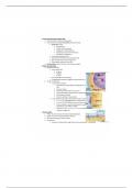
Mobitz type 1 (Wenkebach): PR progressively elongates until
there is failure of conduction of an atrial beat (then the cycle CARDIAC CONDUCTION CIRCUIT
1st and 2nd AV block may be repeats) SA node
caused by: ↑vagal tone/athletes,
coronary artery disease, Mobitz type 2: constant normal PR with occasional dropped
myocarditis, acute rheumatic beats
carditis, digoxin toxicity, or
electrolyte disturbances. 2nd degree AV block with 2:1/3:1/4:1 block: alternate AV node
Bundle of His LBB
conducted and non-conducted atrial beats (P:QRS) RBB
Anterior
3rd degree (complete) AV block: complete dissociation between p waves fascicle
and QRS complexes. Normal atrial beats which are not conducted to
ventricles resulting in ventricles self-depolarising at a much slower rate Posterior
‘ventricular escape rhythm’. fascicle
3rd degree AV block is caused by
fibrosis around Bundle of His
(may be caused by ischaemia,
congential, idiopathic, aortic
stenosis, or trauma) or block of
both bundle branches.
© 2014 Dr Christopher Mansbridge at www.OSCEstop.com, a source of free OSCE exam notes for medical students’ finals OSCE revision