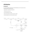
H0: Introduction
Different imaging techniques based on waves:
• X-ray: radiography and CT
• Radiowaves: MRI
• 𝜸-wave: PET & SPECT
• Visible light: optical imaging & bioluminescence imaging
• Soundwaves: US
Different imaging techniques visualize different processes
⤷ The techniques are complementary and provide different information
⤷ overlay of the techniques provides a better understanding of the body (ex. MRI and PET)
There needs to be a compromise between sensitivity and resolution
• The lower the spatial resolution, the better
⤷ you can see more in detail
• The higher the molecular sensitivity, the better
⤷ you want to detect the smallest change
• The ‘ideal’ tool does not exist!
H1: Magnetic Resonance Imaging (MRI)
1.1 General
• Big magnet where the subject is put into
o Patient table that goes in and out of the scanner
o Magnet: sort of a cylinder
o Radiofrequency coils around the area of interest
• MRI exists of 4 categories: Nuclear – Magnetic – Resonance - Imaging
• Most of the techniques were developed for humans, which means that the resolutions of the human machines
are not good enough for the smaller brains, so the image will never be as detailed as in humans
o Clinical scanners: 1-3 Tesla, resolution 1-3 mm
o Preclinical scanners: solution = increasing the magnetic strength (Tesla) to 7-9.4 Tesla, resolution 125-70
µm (the resolution should go to nm, but that is not possible nowadays)
1.2 Nuclear Magnetic Resonance imaging
• Nuclear = signal is based on atomic nuclei
o Usage of protons (+) and neutrons (0) with spin ½ (rotation)
o Hydrogen rotates ➝ creating a magnetic momentum by reversing the field
• Spinning protons: positive charged sphere representing an electrical loop around the axis of rotation
o Electrical current creates a magnetic field (right hand rule)
o So a proton is a sort of small magnet with a given magnetic momentum µ
o Not all nuclei can be used, as pairs tend to cancel each other
⤷ we need to use atoms with an odd number of protons or neutrons
o We are basically measuring hydrogen with NMR (high abundance in the body)
⤷ other atoms (13C, 14N, 23Na, 31P, 19F) will barely give a signal and other important biological molecules
such as 12C and 16O cannot be seen because they don’t have magnetic resonance
1
, 1.3 MRI signal
• Protons in tissue are randomly spinning in different orientations so there’s no magnetic field
• Subject in magnetic field with given direction (B0): protons tend to align with this magnetic field = equilibrium
o Protons in magnetic field will be hindered in their spin orientation
⤷ they will align upwards the magnetic field (low energy state) and downwards (higher energy state)
with most of the protons aligned upwards
⤷ because they are all aligned in the same direction, they will create a signal: longitudinal magnetization
• The protons will always have some kind of angle, so will not be perfectly aligned with the B0 (depending on how
strong the magnetic field is) → they will spin around the orientation of the magnetic field
o Given by the Larmor frequency: F = 𝜸 x B0 (frequency of this movement is proportional to B0 with
gamma, the angle at which the spin is present)
• When given a RF pulse by an antenna (typically 90°), you will displace the equilibrium and the protons will flip
and go towards an higher energy state
o The protons have the tendency to go towards the orientation of the pulse
o Longitudinal magnetization is lost, and at the same time you will get transverse magnetization
o This is what you can detect with the RF antenna
⤷ proportionally related to the amount of protons (hydrogen atoms) that are present in the tissue
⤷ more atoms = proton density higher = signal higher
• All of the spins will want to return to the lower energy state: relaxation process
o to do so they will have to release energy in order to eventually realign back with B0
o this can happen in 2 ways (T1 and T2 are different in different tissues)
1. Spin lattice T1 relaxation: given by the release of energy from these protons to the surrounding
tissue (often as heat) => automatically go back to longitudinal magnetization (equilibrium)
2. Spin-spin T2 relaxation: given by the interaction of given protons to each other. When we are in
transverse magnetization, we have many protons in that orientation and the same direction, but
because they are all positively charged, they will tend to repel each other => little by little they will
stop having an in-phase orientation and will get disorientation => total summation will become zero
(this has nothing to do with energy!)
• Extra concepts:
o TR (repetition time) is the time between two consecutive RF pulses (to keep measuring, the more times
you do it, the more information you can get)
o TE (echo time) is the time between RF pulse (irridation) and receiving the signal from the transverse
magnetization decrease
o By correctly choosing TR and TE of successive RF pulses, your contrast can be either T1 or T2 weighted
o Signal is based on the number of hydrogen atoms. Tissue: 60-80% water ➝ relatively constant
▪ It would require contrast enhancement => change water content ((de)hydration)
▪ Impractical approach
2