Pig Kidney Dissection
Introduction:
In mammals the two kidneys are found close to the back wall of the abdominal cavity on either side of the
vertebral column. Their functions include the filtering of metabolic wastes such as urea and salts from the
blood, and maintaining a balance of water, salts and pH.
Purpose:
To examine the external and internal structure of a kidney and to relate structure to function.
Materials:
Sheep, pig or cow kidney Safety glasses Disposable gloves
Dissecting tray Scalpel Plastic bags (to store or dispose of specimen)
Safety:
● Extreme care must be taken when using dissecting instruments, particularly scalpels
● Dispose of all materials as instructed by your teacher, and clean your work area thoroughly
Procedure
1. Place the kidney on its side on the dissection board and carefully remove the fat from around the kidney.
2. Observe the external features of the kidney. The renal capsule is a smooth, semi-transparent membrane
that is tightly bound to the outer surface of the kidney. Identify and remove the renal capsule.
3. Under the surface of the renal capsule is the surface of the renal cortex. Locate the area where the renal
blood vessels and the ureter are attached to the kidney.
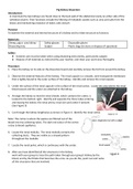
4. Arrange the kidney so that the renal sheath, which contains the ureter, is
located to the bottom right. Identify and separate the three tubes entering
and leaving the kidney: the renal artery, renal vein and ureter if present.
(See Figure 3)
5. Cut through the kidney lengthwise as shown in Figure 4. Identify the renal cortex.
Note: The cortex is where the wastes are filtered out of the
blood into tiny collecting tubes. The dark red colour of the cortex
is due to blood capillaries.
6. Locate the renal medulla. The renal medulla contains the
collecting ducts. They are visible as a striped pattern
throughout the medulla.
7. Locate the renal pelvis, which is continuous with the ureter.
8. After you have identified all the structures in the kidney,
work with your group to trace the path taken through your group’s kidney by the
blood, and by the filtrate that becomes the urine. As you do this, point out and name
all the structures that are involved.
Introduction:
In mammals the two kidneys are found close to the back wall of the abdominal cavity on either side of the
vertebral column. Their functions include the filtering of metabolic wastes such as urea and salts from the
blood, and maintaining a balance of water, salts and pH.
Purpose:
To examine the external and internal structure of a kidney and to relate structure to function.
Materials:
Sheep, pig or cow kidney Safety glasses Disposable gloves
Dissecting tray Scalpel Plastic bags (to store or dispose of specimen)
Safety:
● Extreme care must be taken when using dissecting instruments, particularly scalpels
● Dispose of all materials as instructed by your teacher, and clean your work area thoroughly
Procedure
1. Place the kidney on its side on the dissection board and carefully remove the fat from around the kidney.
2. Observe the external features of the kidney. The renal capsule is a smooth, semi-transparent membrane
that is tightly bound to the outer surface of the kidney. Identify and remove the renal capsule.
3. Under the surface of the renal capsule is the surface of the renal cortex. Locate the area where the renal
blood vessels and the ureter are attached to the kidney.
4. Arrange the kidney so that the renal sheath, which contains the ureter, is
located to the bottom right. Identify and separate the three tubes entering
and leaving the kidney: the renal artery, renal vein and ureter if present.
(See Figure 3)
5. Cut through the kidney lengthwise as shown in Figure 4. Identify the renal cortex.
Note: The cortex is where the wastes are filtered out of the
blood into tiny collecting tubes. The dark red colour of the cortex
is due to blood capillaries.
6. Locate the renal medulla. The renal medulla contains the
collecting ducts. They are visible as a striped pattern
throughout the medulla.
7. Locate the renal pelvis, which is continuous with the ureter.
8. After you have identified all the structures in the kidney,
work with your group to trace the path taken through your group’s kidney by the
blood, and by the filtrate that becomes the urine. As you do this, point out and name
all the structures that are involved.