4.1: Neuromuscular function
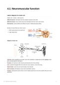
Label a diagram of a motor unit
[ Motor unit = muscle + motor neuron ]
Afferent neurons: information from the receptors (sensors) to the CNS
Efferent neurons: motor neurons that carry informations from the CNS to the muscle
Interneurons: connect afferent and efferent neurons. Inhibitory/excitatory effect
Number of muscle fibers per motor neuron:
Small: great precision but small force
Large: large forces
Diagram of motor unit
Cell body: where organelles are located. most of the cytoplasm is located there and the nucleus is also
there (contains genetic information)
Dendrites: link neurons to other neurons and transmits info to the cell body
Axon: cylindrical elongated fiber that carries the nerve impulse. can be 20cm long.
Myelin sheath: covers the axon and increases nerve impulse
Motor end plate: joint between neuron and muscle/other neurons.
Synapse: small junction across where a nerve impulse passes from one cell to another (they don’t touch
eachother)
4.1: Neuromuscular function 1
, Muscle structure and sacromere
[ skeletal muscle - fascicle - muscle fiber - myofibril ]
Skeletal muscles are formed by a group of muscle cells (muscle fiber) that are packed into bundles
(fascicle). Muscle fibers are formed by overlapping proteins (actin and myosin) in a structure called
myofibril.
Basic structure of myofibril: sacromere
Thin filament: actin
Thick filament: myosin
Actin attaches to the Z-line at one end and
myosin is in the middle.
sacromere
H-zone: region of myosin only (lightest one)
Z-line: perpendicular stripe of t-tubules where
actin attaches
*sacromere is what whe find between two Z-
lines
A-band: total lenght of myosin
I-band: region of actin only
M-line: perpendicular line of proteins in the middle
During muscle contraction
actin filaments run along myosin filaments towards M-line and shortens muscle fiber
4.1: Neuromuscular function 2