A&P 1 101 Module 4 Skeletal system TEST; Portage Learning
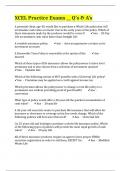
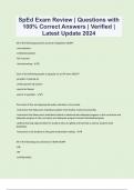
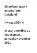
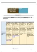
A&P 1 101 Module 4 Skeletal system TEST Portage Learning A&P 1 101 Module 4 Skeletal system TEST; Portage Learning Exam Page 1 Answer the following five questions: 1. Which of the following statements is TRUE concerning the function bones? A. Sesamoid bones are large and thin. B. Short bones have an irregular structure C. The fontanelles are an example of sesamoid bone. D. Vertebrae are an example of irregular bone. D 2. Which of the following statements is FALSE concerning bones? A. Bones are a storage site for phosphorus and calcium. B. A tuberosity is a projection for a tendon or ligament attachment. C. A process is a depression found in bone. D. Bones are completely smooth surfaces. E. C. and D. are false E 3. Which of the following statements is FALSE about the skeletal system? A. The two main divisions of the skeletal system are: axial and appendicular. B. The two main divisions of the skeletal system are: vertebral and thoracic. C. The axial skeleton lies along the midline of the body. D. The vertebral division of the skeleton is included in the appendicular skeleton. E. B. and D. are false F. A. and C are false E 4. The skull is formed by bones; the facial skeleton contains bones. A. 30; 10 B. 22; 14 C. 10; 5 D. 15; 7 B 5. What bone is highlighted in blue in the figure below? (superior/internal view) Sphenoid bone Answer Key Answer the following five questions: 1. Which of the following statements is TRUE concerning the function bones? A. Sesamoid bones are large and thin. B. Short bones have an irregular structure C. The fontanelles are an example of sesamoid bone. D. Vertebrae are an example of irregular bone. D. Vertebrae are an example of irregular bone. 2. Which of the following statements is FALSE concerning bones? A. Bones are a storage site for phosphorus and calcium. B. A tuberosity is a projection for a tendon or ligament attachment. C. A process is a depression found in bone. D. Bones are completely smooth surfaces. E. C. and D. are false E. C. and D. are false 3. Which of the following statements is FALSE about the skeletal system? A. The two main divisions of the skeletal system are: axial and appendicular. B. The two main divisions of the skeletal system are: vertebral and thoracic. C. The axial skeleton lies along the midline of the body. D. The vertebral division of the skeleton is included in the appendicular skeleton. E. B. and D. are false F. A. and C are false E. B. and D. are false B. 22; 14 5. What bone is highlighted in blue in the figure below? (superior/internal view) Sphenoid bone Copyright © 2020 Portage Learning. All Rights Reserved. Exam Page 2 1. Label the following bones of the skeleton from the figure below: 2:Nasal bone 4:zygomatic bone 6:inferior nasal concha 7:maxilla 8: mandible Answer Key 1. Label the following bones of the skeleton from the figure below: 4: Zygomatic bone 6: Inferior nasal concha 7: Maxilla 8: Mandible Copyright © 2020 Portage Learning. All Rights Reserved. Exam Page 3 Answer the following three questions: 1. Label the following vertebrae as: A= Cervical B= Thoracic C= Lumbar c 2. Label the following vertebrae as: A= Cervical B= Thoracic C= Lumbar b 3. What is the name of the foramina in the figure below? 1:Carotid canal 2:external acoustic meatus 3:stylomastoid foramen -3.0 points Answer Key Answer the following three questions: 1. Label the following vertebrae as: A= Cervical B= Thoracic C= Lumbar A (Cervical) 2. Label the following vertebrae as: A= Cervical B= Thoracic C= Lumbar B (Thoracic) 2: Carotid canal 3: External acoustic meatus Copyright © 2020 Portage Learning. All Rights Reserved. Exam Page 4 1. Label the following bone landmarks: A:head C:lesser tubercle F:deltoid tuberosity I:capitulum K:medial epicondyle Answer Key 1. Label the following bone landmarks: A: Head (optional: of the humerus) C: Lesser tubercle F: Deltoid tuberosity I: Capitulum K: Medial epicondyle Copyright © 2020 Portage Learning. All Rights Reserved. Exam Page 5 Answer the following three questions: 1. Which of the following statements is TRUE about the scapula? A. The clavicle connects to the scapula anteriorly near the midline of the human body. B. The medial border of the scapula connects directly to the neck of the scapula. C. The subscapular fossa is located on the anterior side of the scapula. D. The scapula articulates with the clavicle at the neck of the scapula. c 2. Which of the following statements is FALSE about the humerus? A. The trochlea articulates with the ulna. B. The head of the humerus articulates with the ulna. C. The capitulum articulates with the radius. D. The medial epicondyle is the prominent bone landmark of the medial side of the elbow (in anatomical position). E. A. and B. are false e 3. What two bones meet at the glenohumeral joint? In your own words, why is this joint prone to dislocation? The humerus and scapula. The joint is prone to dislocation because it is held in place primarly by muscular and ligament attachment with very little stability. Answer Key Answer the following three questions: 1. Which of the following statements is TRUE about the scapula? A. The clavicle connects to the scapula anteriorly near the midline of the human body. B. The medial border of the scapula connects directly to the neck of the scapula. C. The subscapular fossa is located on the anterior side of the scapula. D. The scapula articulates with the clavicle at the neck of the scapula. C. The subscapular fossa is located on the anterior side of the scapula. 2. Which of the following statements is FALSE about the humerus? A. The trochlea articulates with the ulna. B. The head of the humerus articulates with the ulna. C. The capitulum articulates with the radius. D. The medial epicondyle is the prominent bone landmark of the medial side of the elbow (in anatomical position). E. A. and B. are false B. The head of the humerus articulates with the ulna. (The head of the humerus articulates with the scapula) 3. What two bones meet at the glenohumeral joint? In your own words, why is this joint prone to dislocation? Humerus and scapula The structure of the shoulder permits movement of the arm in almost any direction but provides little stability. The glenohumeral joint is prone to dislocation because it is held in place primarily by muscular and ligament attachment with very little bony stability. Copyright © 2020 Portage Learning. All Rights Reserved. Exam Page 6 1. Label the bones in the figure below: A:pisiform B:hamate C:capitate D:trapezoid E: trapezium 2. Label the bones in the figure below: A:cuboid C:intermediate cuneiform D:medial cuneiform F:navicular G:calcaneus Answer Key 1. Label the bones in the figure below: A: Pisiform B: Hamate C: Capitate D: Trapezoid E: Trapezium Intermediate cuneiform D: Medial Cuneiform F: Navicular G: Calcaneus Copyright © 2020 Portage Learning. All Rights Reserved. Exam Page 7 Answer the following five questions: 1. Yellow bone marrow: A. is found primarily in long bones. B. is found primarily in short and flat bones. C. is found primarily in newborns, not adults. D. produces red blood cells. a 2. The diaphysis of a bone: A. is found at the ends of long bones. B. contains the articular cartilage at joint articulations. C. contains the proximal epiphysis. D. is the center length of a bone. E. both A. and C. c 3. Compact bone: A. forms the exterior of bones. B. forms the interior of bones. C. is lighter than spongy bone. D. contains numerous bars and plates with irregular spaces. E. both B. and D. a 4. Intramembranous ossification is the formation of from A. a growth plate; the center of a bone. B. long bones; hyaline cartilage. C. flat bones; connective tissue. D. a primary ossification center; a cartilaginous disc. E. both A. and C. : c 5. What term best describes the type of fracture pictured below? traverse -4.0 points Answer Key Answer the following five questions: 1. Yellow bone marrow: A. is found primarily in long bones. B. is found primarily in short and flat bones. C. is found primarily in newborns, not adults. D. produces red blood cells. A. is found primarily in long bones. 2. The diaphysis of a bone: A. is found at the ends of long bones. B. contains the articular cartilage at joint articulations. C. contains the proximal epiphysis. D. is the center length of a bone. E. both A. and C. D. is the center length of a bone. 3. Compact bone: A. forms the exterior of bones. B. forms the interior of bones. C. is lighter than spongy bone. D. contains numerous bars and plates with irregular spaces. E. both B. and D. A. forms the exterior of bones. 4. Intramembranous ossification is the formation of from : A. a growth plate; the center of a bone. B. long bones; hyaline cartilage. C. flat bones; connective tissue. D. a primary ossification center; a cartilaginous disc. E. both A. and C. C. flat bones; connective tissue 5. What term best describes the type of fracture pictured below? Greenstick (the bone is broken, but not all the way across) Copyright © 2020 Portage Learning. All Rights Reserved. Exam Page 8 Answer the following two questions: 1. A patient has a diagnosis of osteoporosis. (1) In your own words, describe this diagnosis and (2) What type of bone cell would they be lacking? Explain your answer. Osteoporosis is a bone disease that causes bone tissue to degenerate faster than it can be replaced. This causes pain and more like to a fracture. The bone cell that would be lacking is osteoblast.Osteoblast is responsible for bone rapair. 2. Your patient has back pain due to a herniated disc. (1) In your own words explain what it means to have a herniated disc. (2) As reviewed in the module, discuss one treatment option to address your patient’s pain. 1) A herniated disc is an injury to the intervertebral disc, where the center portion of the disc bulges into the verebral foramen causing pain. 2) Physical therapy for strengthening to support back ligaments Answer Key Answer the following two questions: 1. A patient has a diagnosis of osteoporosis. (1) In your own words, describe this diagnosis and (2) What type of bone cell would they be lacking? Explain your answer. (1) Osteoporosis is a bone tissue disease. When bone tissue degenerates faster than is replaced, the bones become weak. Brittle bones cause increased pain and are more likely to fracture. (2) They would have decreased osteoblasts which are responsible for bone repair. The bone repair would be unable to keep up with the ongoing breakdown of bone which is done by the work of osteoblasts. 2. Your patient has back pain due to a herniated disc. (1) In your own words explain what it means to have a herniated disc. (2) As reviewed in the module, discuss one treatment option to address your patient’s pain. (1) A herniated disc is an injury to the intervertebral disc, where the center portion of the disc bulges into the vertebral foramen, causing pain. (2) Explanation of 1- Physical therapy for strengthening to support back ligaments. OR 2- Surgery to fuse two vertebrae together. Copyright © 2020 Portage Learning. All Rights Reserved. Exam Page 9 1. Matching: Match the joint with the correct joint classification (A-F). *NOTE: Some joints may fall into more than one category. Mark all that apply. A= Fibrous B= Cartilaginous C= Synovial D= Hinge E= Ball-and-Socket F=Saddle 1. Elbow joint c, d 2. Thumb joint f, c 3. Hip joint e 4. Vertebral joint b 5. Cranial joints a -1.0 points Answer Key 1. Matching: Match the joint with the correct joint classification (A-F). *NOTE: Some joints may fall into more than one category. Mark all that apply. A= Fibrous B= Cartilaginous C= Synovial D= Hinge E= Ball-and-Socket F=Saddle 1. Elbow joint C, D (Synovial, Hinge) 2. Thumb joint C, F (Synovial, Saddle) 3. Hip joint C, E (Synovial, Ball and socket) 4. Vertebral joint B (Cartilaginous) 5. Cranial joints A (Fibrous) Copyright © 2020 Portage Learning. All Rights Reserved. Exam Page 10 Answer the following three questions: 1. Name the ligament highlighted in blue in the figure below: anterior sacroiliac ligament 2. Name the ligament highlighted in blue in the figure below: Medial collateral/tibial ligament 3. Name the ligament highlighted in blue in the figure below: acromiovlavicular ligament Answer Key Answer the following three questions: 1. Name the ligament highlighted in blue in the figure below: Anterior sacroiliac ligament MCL (Medial collateral/tibial ligament) 3. Name the ligament highlighted in blue in the figure below: Acromioclavicular ligament Copyright © 2020 Portage Learning. All Rights Reserved.
Escuela, estudio y materia
- Institución
-
Portage Learning
- Grado
-
A&P 1 101
Información del documento
- Subido en
- 19 de octubre de 2022
- Número de páginas
- 40
- Escrito en
- 2022/2023
- Tipo
- Examen
- Contiene
- Preguntas y respuestas
Temas
-
aampp 1 101 module 4 skeletal system test portage learning
-
aampp 1 101 module 4 skeletal system test portage learning aampp 1 101 module 4 skeletal system test portage learning
Documento también disponible en un lote