The Muscles of Mastication
The muscles of mastication are associated with movements of the jaw
(temporomandibular joint). They are one of the major muscle groups in the head –
the other being the muscles of facial expression. There are four muscles:
Masseter
Temporalis
Medial pterygoid
Lateral pterygoid
The muscles of mastication develop from the first pharyngeal arch. Thus, they are
innervated by a branch of the trigeminal nerve (CN V), the mandibular nerve.
In this article, we shall look at the anatomy of the muscles of mastication – their
attachments, actions, and innervation.
(NB: It is important to note that all the muscles mentioned here are bilateral
structures).
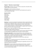
Masseter
The masseter muscle is the most powerful muscle of mastication. It is quadrangular
in shape and has two parts: deep and superficial.
The entirety of the muscle lies superficially to the pterygoids and temporalis,
covering them.
Attachments: The superficial part originates from maxillary process of the
zygomatic bone. The deep part originates from the zygomatic arch of the temporal
bone. Both parts attach to the ramus of the mandible.
Actions: Elevates the mandible, closing the mouth.
Innervation: Mandibular nerve (V3).
The muscles of mastication are associated with movements of the jaw
(temporomandibular joint). They are one of the major muscle groups in the head –
the other being the muscles of facial expression. There are four muscles:
Masseter
Temporalis
Medial pterygoid
Lateral pterygoid
The muscles of mastication develop from the first pharyngeal arch. Thus, they are
innervated by a branch of the trigeminal nerve (CN V), the mandibular nerve.
In this article, we shall look at the anatomy of the muscles of mastication – their
attachments, actions, and innervation.
(NB: It is important to note that all the muscles mentioned here are bilateral
structures).
Masseter
The masseter muscle is the most powerful muscle of mastication. It is quadrangular
in shape and has two parts: deep and superficial.
The entirety of the muscle lies superficially to the pterygoids and temporalis,
covering them.
Attachments: The superficial part originates from maxillary process of the
zygomatic bone. The deep part originates from the zygomatic arch of the temporal
bone. Both parts attach to the ramus of the mandible.
Actions: Elevates the mandible, closing the mouth.
Innervation: Mandibular nerve (V3).