
• ¿Documento equivocado? Cámbialo gratis
  • Escrito por estudiantes que aprobaron
  • Inmediatamente disponible después del pago
  • Leer en línea o como PDF
Vender
¿Dónde estudias?
Tu idioma
Document preview thumbnail
Vista previa 1 fuera de 2 páginas
Examen

Microscope Lab Worksheet

Document preview thumbnail
Vista previa 1 fuera de 2 páginas

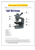
Microscope Lab Worksheet Name: I. Please label the microscope diagram below. A. Objective lens B. Stage C. Diaphragm D. Light source E. Ocular or eyepiece F. Stage clips G. Course focus knob H. Fine focus knob II. Microscope Comparison (write a single paragraph) A light microscope, as it sounds, uses light to illuminate specimens and a glass lens is used to magnify. An electron microscope uses beams of electrons to illuminate specimens and a magnetic lens is used to magnify. The resolution of image that you get from the electron microscope is about 0.01 nanometers while a light microscope is only 3 micrometers. With that being said then as you can imagine a cell under an electron microscope would even be able to see the nucleus. The electron microscope however cannot be used on live specimens as the electrons can be harmful to living things, so then a light microscope would be of use. The type of microscope used for the skin cell images for this assignment is fairly obvious. The left image is the light microscope where it’s still fairly detailed but you see multiple cells while the right image with the electron microscope shows the detail of an individual cell and I believe you can even see it as if it were somewhat three dimensional or the texture of the surface, however you cannot see it in color. With the light microscope you can see pigments of color or you can dye the specimen.

Vista previa del contenido

Microscope Lab Worksheet
Name:

I. Please label the microscope diagram below.


A. Objective lens
B. Stage
C. Diaphragm
D. Light source
E. Ocular or eyepiece
F. Stage clips
G. Course focus knob
H. Fine focus knob


II. Microscope Comparison (write a single paragraph)

A light microscope, as it sounds, uses light to illuminate specimens and a glass lens is used to magnify. An
electron microscope uses beams of electrons to illuminate specimens and a magnetic lens is used to
magnify. The resolution of image that you get from the electron microscope is about 0.01 nanometers while
a light microscope is only 3 micrometers. With that being said then as you can imagine a cell under an

1 Microscope Lab Worksheet

Información del documento

Subido en
29 de marzo de 2022
Número de páginas
2
Escrito en
2022/2023
Tipo
Examen
Contiene
Preguntas y respuestas
$12.99

¿Documento equivocado? Cámbialo gratis Dentro de los 14 días posteriores a la compra y antes de descargarlo, puedes elegir otro documento. Puedes gastar el importe de nuevo.
Escrito por estudiantes que aprobaron
Inmediatamente disponible después del pago
Leer en línea o como PDF

Seller avatar
Los indicadores de reputación están sujetos a la cantidad de artículos vendidos por una tarifa y las reseñas que ha recibido por esos documentos. Hay tres niveles: Bronce, Plata y Oro. Cuanto mayor reputación, más podrás confiar en la calidad del trabajo del vendedor.
NURSEREP
4.7
(328)
Vendido
602
Seguidores
424
Artículos
2671
Última venta
1 semana hace


Por qué los estudiantes eligen Stuvia

Creado por compañeros estudiantes, verificado por reseñas

Calidad en la que puedes confiar: escrito por estudiantes que aprobaron y evaluado por otros que han usado estos resúmenes.

¿No estás satisfecho? Elige otro documento

¡No te preocupes! Puedes elegir directamente otro documento que se ajuste mejor a lo que buscas.

Paga como quieras, empieza a estudiar al instante

Sin suscripción, sin compromisos. Paga como estés acostumbrado con tarjeta de crédito y descarga tu documento PDF inmediatamente.

Student with book image

“Comprado, descargado y aprobado. Así de fácil puede ser.”

Alisha Student

Preguntas frecuentes
