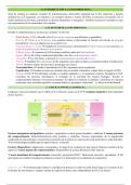
1) Discuss the statement that ‘mitochondria are the critical control centres of apoptosis’.
Cellular apoptosis can be induced intrinsically or extrinsically either via internal stimuli such as DNA
damage or metabolic stress or extracellular ligands such as FasL. However, the statement that
‘mitochondria are the critical control centres of apoptosis’ provides an area for further research into
the key controls of apoptosis. It is thought that there are three main contenders for the role of
critical control centres, including caspases, BCL-2 proteins, and the mitochondria. In this essay all
three elements can be considered the critical control centre for apoptosis will be discussed.
Caspases
There are two groups of caspases involved in apoptosis, initiator caspases (caspase-2, -8, -9 and -10)
which are primarily responsible for the initiation of the apoptotic pathway and effector caspases
(caspase-3, -6 and -7) which are responsible in the actual cleavage of cellular components during
apoptosis. Pro-caspase 9 is the main caspase which triggers the further degradation of the cell. Once
the outer mitochondrial matrix (OMM) has been permeabilised, through the process of MOMP
(mitochondrial outer membrane permeabilisation) in which pro-apoptotic Bcl-2 proteins, BAX and
BAK are recruited to OMM and oligomerise to form pores in the OMM facilitating the release of
cytochrome C. Cytochrome C activates Apaf-1 by hydrolysis of bound dATP to dADP. Apaf-1
undergoes a conformational change allowing it to bind to pro-caspase 9 via CARD (caspase
recruitment domain) forming the apoptosome. Pro-caspase 9 is activated by the apoptosome and
caspase 9 can the cleave and thereby activate executioner pro-caspases. However caspase 9’s ability
to cleave and activate pro-caspase 3, 6 and 7 has been shown the be dependent on the apoptosome,
according to a Boatright et al (2003) the crystal structure of the caspase-9 dimer shows that only one
of the two subunits in the dimer is catalytically active. Yin et al (2006) discovered that caspase-9 was
a completely unique caspase due to its mechanism of activation and its affinity for pro-caspase 3,
although other caspases such as caspase-8 can directly activate its substrates without the
requirement to be bound to an accelerator, caspase 9 when associated with the apoptosome it is
termed the caspase 9 holoenzyme (C9holo) (Rodriguez and Lazebnik, 1999). Without the activation
of effector caspases, regardless of the activation being intrinsic or extrinsic, apoptosis would not
occur without the activity of caspases 9, 3, 6 and 7.
BCL-2 proteins
Pro-apoptotic proteins BAX and BAK are part of the BCL-2 family. Included in the family are BH-3
only proteins an example of which is PUMA, the p53 upregulated modulator of apoptosis. PUMA
acts as a sensor to the accumulation of p53 when apoptosis is initiated and acts to inhibit anti-
apoptotic signals such as BCL-2 and BCL-X. As stated, BAX and BAK are recruited the OMM to
oligomerise and form pores in the mitochondrial membrane which allow the mitochondrial contents
to enter the cytosol including cytochrome C. This is an essential part of apoptosis, once the
mitochondria have begun degrading there is no returning, the cell must commit to apoptosis from
that point forwards. However, the actual mechanism for activation of BAX/BAK is still unknown.
There are theories on the mechanism but none of which have been proved yet. Luo, O’Neill, and
Huang (2020) discussed some theories based on BH-3 only mediated activation of BAX and BAK,
including direct activation which suggests that activators directly bind to and activate BAX/BAK when
they are not isolated by anti-apoptotic BCL-2 proteins. Another theory suggested an indirect
mechanism which suggests that BAX/BAK are constitutively active but are inhibited by the anti-
apoptotic BCL-2 proteins, yet, during apoptosis the BH3-only proteins bind to the anti-apoptotic BCL-