Lecture 3: Axial Skeleton and the Forelimb
Learning Objectives
1) List the bones of the axial skeleton and identify their specific features
2) List and identify the bones of the forelimb and list some of the most
relevant species variations
Axial Skeleton - The Framework
1. Skull (and the hyoid apparatus)
2. Spinal column (made up by the vertebrae)
3. Ribs
4. Sternum (made by individual sternebrae)
Appendicular Skeleton
a. Forelimb
● Clavicle (cats only) - very rare in dogs
● Scapula - shoulder blade equivalent
● Humerus
● Radius and ulna
● Carpal bones (radiocarpal, ulnarcarpal, accessory and I, II, III, IV and
V)
● Metacarpal bones (I-V)
● Phalanges (within the digit), 1, 2 and 3. Each of the three digits has
three phalanges (except digit 1, the dew claw)
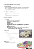
Axial Skeleton
1. Skull
● Cranium - subdivided into other
bones
● Maxilla
● Mandible
● Hyoid apparatus
○ Attached in the
caudo-ventral aspect of the cranium which holds the windpipe in
place
Skull Functions
1. To protect and house the brain
, 2. Housing for the sense organs i.e. the nose, ear, eye and tongue
3. To provide housing for the mandible (lower jaw)
4. To provide attachment for the hyoid apparatus
5. To attach the facial muscles
6. To host teeth
7. To attach the larynx (part of the respiratory system, linked to the hyoid
apparatus)
Bones in the Skull
a. Parietal (dorso-lateral walls)
b. Occipital (caudo-ventral surface)
c. Temporal (caudo-lateral wall) that link to the zygomatic bones through the
zygomatic arch
d. Lacrimal (rostro-medial to the zygomatic arch)
e. Palatine bone (ventro-medial to the zygomatic arch)
f. Sphenoid bones (caudo-medial to the zygomatic arch)
g. Pterygoid (ventral to the palatine and the sphenoid)
h. Basisphenoid (caudal to the pterygoid)
i. Incisive (rostral and ventral to the nasal bone)
j. Nasal (dorsal to incisive and rostral to the maxilla)
Skull
● Most joints are fibrous (called sutures)
● Temporomandibular joint is synovial (TMJ)
● Each mandible joins by mandibular
symphysis
, Bony landmarks of clinical importance
● Foramen magnum
○ Large opening in the occipital bone bone of
the skull
○ Passageway for spinal cord - an extension
of the medulla oblongata
● Occipital condyles
● Tympanic bulla
● Zygomatic arch
● Temporo-mandibular joints (TMJ)
● Orbit
● Occipital crest
● Mandible: ramus, angle of jaw and
body
Nasal Passages and Maxilla
● The maxilla, palatine and incisive bones form the hard palate (maxilla +
palatine + incisive bones = hard palate)
● Nasal chamber is divided by the nasal septum
● Each chamber contains turbinate bones which are called conchae - look like a
seashell
● Caudal of conchae: ethmoidal labyrinth - full of blood, responsible for
nosebleeds, don’t want to poke it when doing an endoscopy
Learning Objectives
1) List the bones of the axial skeleton and identify their specific features
2) List and identify the bones of the forelimb and list some of the most
relevant species variations
Axial Skeleton - The Framework
1. Skull (and the hyoid apparatus)
2. Spinal column (made up by the vertebrae)
3. Ribs
4. Sternum (made by individual sternebrae)
Appendicular Skeleton
a. Forelimb
● Clavicle (cats only) - very rare in dogs
● Scapula - shoulder blade equivalent
● Humerus
● Radius and ulna
● Carpal bones (radiocarpal, ulnarcarpal, accessory and I, II, III, IV and
V)
● Metacarpal bones (I-V)
● Phalanges (within the digit), 1, 2 and 3. Each of the three digits has
three phalanges (except digit 1, the dew claw)
Axial Skeleton
1. Skull
● Cranium - subdivided into other
bones
● Maxilla
● Mandible
● Hyoid apparatus
○ Attached in the
caudo-ventral aspect of the cranium which holds the windpipe in
place
Skull Functions
1. To protect and house the brain
, 2. Housing for the sense organs i.e. the nose, ear, eye and tongue
3. To provide housing for the mandible (lower jaw)
4. To provide attachment for the hyoid apparatus
5. To attach the facial muscles
6. To host teeth
7. To attach the larynx (part of the respiratory system, linked to the hyoid
apparatus)
Bones in the Skull
a. Parietal (dorso-lateral walls)
b. Occipital (caudo-ventral surface)
c. Temporal (caudo-lateral wall) that link to the zygomatic bones through the
zygomatic arch
d. Lacrimal (rostro-medial to the zygomatic arch)
e. Palatine bone (ventro-medial to the zygomatic arch)
f. Sphenoid bones (caudo-medial to the zygomatic arch)
g. Pterygoid (ventral to the palatine and the sphenoid)
h. Basisphenoid (caudal to the pterygoid)
i. Incisive (rostral and ventral to the nasal bone)
j. Nasal (dorsal to incisive and rostral to the maxilla)
Skull
● Most joints are fibrous (called sutures)
● Temporomandibular joint is synovial (TMJ)
● Each mandible joins by mandibular
symphysis
, Bony landmarks of clinical importance
● Foramen magnum
○ Large opening in the occipital bone bone of
the skull
○ Passageway for spinal cord - an extension
of the medulla oblongata
● Occipital condyles
● Tympanic bulla
● Zygomatic arch
● Temporo-mandibular joints (TMJ)
● Orbit
● Occipital crest
● Mandible: ramus, angle of jaw and
body
Nasal Passages and Maxilla
● The maxilla, palatine and incisive bones form the hard palate (maxilla +
palatine + incisive bones = hard palate)
● Nasal chamber is divided by the nasal septum
● Each chamber contains turbinate bones which are called conchae - look like a
seashell
● Caudal of conchae: ethmoidal labyrinth - full of blood, responsible for
nosebleeds, don’t want to poke it when doing an endoscopy