
BIOS 252 / Anatomy & Physiology II with Lab | Lab Practicum Review Chamberlain University | 2026/2027 Edition
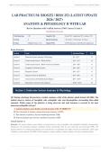
LAB PRACTICUM: BIOS252 / BIOS 252 (LATEST UPDATE
)
ANATOMY & PHYSIOLOGY II WITH LAB
Review Questions with Verified Answers | 100% Correct | Grade A
Chamberlain University
Total Questions 120 Cognitive Mix Recall 25% | Application 50% | Analysis 25%
Sections 8 Special Inclusions 30 Lab ID | 17 Physiology | 9 Clinical
Format Multiple Choice (A-D) Aligned With HAPS Outcomes & A&P II Lab Competencies
Exam Structure
Section Topic Question Range # Qs
Section 1 Endocrine System Anatomy & Physiology Q1 - Q15 15
Section 2 Cardiovascular System - Blood & Heart Q16 - Q33 18
Section 3 Cardiovascular System - Blood Vessels & Circulation Q34 - Q48 15
Section 4 Lymphatic & Immune Systems Q49 - Q60 12
Section 5 Respiratory System Q61 - Q75 15
Section 6 Digestive System & Metabolism Q76 - Q90 15
Section 7 Urinary System & Fluid/Electrolyte Balance Q91 - Q105 15
Section 8 Reproductive Systems & Development Q106 - Q120 15
Section 1: Endocrine System Anatomy & Physiology
Q1: During a histology lab practical, a student examines a slide of the pituitary gland stained with H&E. The
student observes clusters of basophilic cells, acidophilic cells, and chromophobes surrounding thin-walled
sinusoids. Which region of the pituitary is being observed, and what hormone is secreted by the most
numerous acidophilic cell type?
A. Anterior pituitary (pars distalis); growth hormone (GH) *[CORRECT]*
B. Pars intermedia of pituitary; adrenocorticotropic hormone (ACTH)
C. Pars tuberalis of pituitary; thyroid-stimulating hormone (TSH)
D. Posterior pituitary (pars nervosa); antidiuretic hormone (ADH)
Correct Answer: A
Rationale: The anterior pituitary (pars distalis) is characterized by cords of glandular cells (acidophils, basophils, chromophobes)
interspersed with fenestrated sinusoids. Acidophils include somatotrophs (GH, the most numerous) and lactotrophs (PRL). The
posterior pituitary (A) contains unmyelinated axons and pituicytes, not glandular cells. Option D is the pars intermedia, a thin region
of Rathke's pouch remnants. The BIOS 252 lab competency requires students to identify pituitary cell types by staining affinity.
BIOS 252 Lab Practicum Exam | Page 1
,BIOS 252 / Anatomy & Physiology II with Lab | Lab Practicum Review Chamberlain University | 2026/2027 Edition
Q2: Which of the following hormones exerts its effect by binding to an intracellular receptor, forming a
hormone-receptor complex that acts as a transcription factor in the nucleus?
A. Antidiuretic hormone (ADH)
B. Cortisol *[CORRECT]*
C. Epinephrine
D. Insulin
Correct Answer: B
Rationale: Steroid hormones such as cortisol are lipid-soluble and diffuse through the plasma membrane to bind intracellular
receptors; the complex then binds hormone response elements (HREs) on DNA to regulate gene transcription. Insulin (A) and ADH (D)
are peptide/protein hormones that bind surface receptors and use second messengers (tyrosine kinase and cAMP, respectively).
Epinephrine (B) is a catecholamine that binds G-protein-coupled surface receptors. This distinction aligns with HAPS learning
outcomes on hormone mechanism of action.
Q3: A patient presents with a TSH level of 18 mIU/L (normal 0.4-4.0) and a free T4 of 0.4 ng/dL (normal
0.8-1.8). Which pathophysiological mechanism best explains these findings?
A. Primary hyperthyroidism with suppressed TSH
B. Secondary hypothyroidism due to pituitary failure
C. Primary hypothyroidism with loss of negative feedback *[CORRECT]*
D. Tertiary hypothyroidism due to hypothalamic dysfunction
Correct Answer: C
Rationale: Elevated TSH with low T4 indicates primary hypothyroidism: the thyroid gland fails to produce T4, so negative feedback
on the anterior pituitary is lost, causing thyrotrophs to secrete excess TSH. In secondary hypothyroidism (B), both TSH and T4 would
be low due to pituitary dysfunction. In tertiary hypothyroidism (D), low TRH leads to low TSH and low T4. Option A is incorrect
because hyperthyroidism would suppress TSH. This is a classic HAPS-application scenario on hypothalamic-pituitary-thyroid axis
feedback.
Q4: A 42-year-old female presents with central obesity, moon facies, purple striae on the abdomen,
hypertension, and a fasting glucose of 145 mg/dL. Laboratory testing reveals elevated serum cortisol with loss
of the normal diurnal rhythm and elevated ACTH. CT imaging shows bilateral adrenal hyperplasia. What is
the most likely diagnosis and underlying mechanism?
A. Iatrogenic Cushing's syndrome from exogenous glucocorticoids
B. Primary adrenal insufficiency (Addison's disease) from adrenal destruction
C. Primary hyperaldosteronism (Conn's syndrome) from adrenal adenoma
D. Cushing's disease from an ACTH-secreting pituitary adenoma *[CORRECT]*
Correct Answer: D
Rationale: The clinical presentation (central obesity, moon facies, striae, hypertension, hyperglycemia) is classic for cortisol excess.
Elevated ACTH with bilateral adrenal hyperplasia indicates Cushing's DISEASE (pituitary ACTH adenoma), not Cushing's
SYNDROME from exogenous steroids (C, which would suppress ACTH). Addison's disease (A) presents with hypocortisolism,
hyperpigmentation, and hypotension. Conn's syndrome (D) causes hypertension and hypokalemia but not the striae or hyperglycemia
of cortisol excess. This scenario addresses the BIOS 252 clinical correlation competency for endocrine disorders.
BIOS 252 Lab Practicum Exam | Page 2
,BIOS 252 / Anatomy & Physiology II with Lab | Lab Practicum Review Chamberlain University | 2026/2027 Edition
Q5: A student observes a histological section of the adrenal gland under low power. From outer capsule to
medulla, the correct sequence of cortical zones and their primary secretory product is:
A. Zona fasciculata (aldosterone); zona glomerulosa (cortisol); zona reticularis (androgens)
B. Zona glomerulosa (aldosterone); zona fasciculata (cortisol); zona reticularis (androgens) *[CORRECT]*
C. Zona glomerulosa (cortisol); zona fasciculata (aldosterone); zona reticularis (androgens)
D. Zona reticularis (cortisol); zona fasciculata (androgens); zona glomerulosa (aldosterone)
Correct Answer: B
Rationale: The adrenal cortex displays three concentric zones summarized by the mnemonic 'GFR - Glomerulosa, Fasciculata,
Reticularis' corresponding to 'Salt, Sugar, Sex' - aldosterone (mineralocorticoid, regulates Na+/K+), cortisol (glucocorticoid, regulates
metabolism), and adrenal androgens (DHEA). The zona glomerulosa lies just beneath the capsule, the zona fasciculata is the widest
middle layer of columnar cells in straight cords, and the zona reticularis borders the medulla. This histological zoning is a core BIOS
252 lab identification requirement.
Q6: Which hypothalamic-hypophyseal portal vessel relationship correctly describes the vascular connection
that allows hypothalamic releasing and inhibiting hormones to reach the anterior pituitary without first
undergoing systemic dilution?
A. Hypothalamic hormones are released into the third ventricle and reach the anterior pituitary by cerebrospinal fluid
flow
B. Hypothalamic neurons directly synapse on anterior pituitary endocrine cells
C. Primary capillary plexus in the median eminence drains via long portal veins to a secondary plexus in the
anterior pituitary *[CORRECT]*
D. Short portal vessels carry hormones from the posterior pituitary to the anterior pituitary
Correct Answer: C
Rationale: The hypothalamic-hypophyseal portal system consists of a primary capillary plexus in the median eminence (supplied by
superior hypophyseal artery) that drains through long portal veins down the infundibular stalk into a secondary capillary plexus in the
anterior pituitary. This arrangement ensures releasing/inhibiting hormones (TRH, CRH, GHRH, GnRH, dopamine, somatostatin)
reach anterior pituitary cells at high concentration. Hypothalamic neurons do NOT directly synapse on anterior pituitary cells (A);
short portal vessels (C) connect posterior to anterior but are not the main route. CSF transport (D) is not a mechanism for anterior
pituitary regulation.
Q7: A patient with type 1 diabetes mellitus administers recombinant human insulin subcutaneously. By which
molecular mechanism does insulin lower blood glucose in target cells such as skeletal muscle and adipose
tissue?
A. Binding to G-protein-coupled receptors that activate adenylate cyclase, increasing cAMP
B. Binding to ligand-gated ion channels that allow glucose to flow into cells down its concentration gradient
C. Diffusing through the plasma membrane to bind intracellular receptors that regulate gene transcription
D. Binding to a receptor tyrosine kinase that auto-phosphorylates and triggers a cascade promoting GLUT4
transporter translocation to the plasma membrane *[CORRECT]*
Correct Answer: D
Rationale: Insulin, a peptide hormone, binds a transmembrane receptor with intrinsic tyrosine kinase activity. Receptor
autophosphorylation initiates an IRS-PI3K-Akt signaling cascade that triggers translocation of GLUT4 glucose transporters from
intracellular vesicles to the plasma membrane, facilitating glucose uptake. Option A describes glucagon's mechanism. Option C
describes steroid hormone action. Option D is incorrect because glucose transport is via facilitative carriers (GLUT), not ion channels.
This addresses the BIOS 252/HAPS mechanism-of-action competency for peptide hormones.
BIOS 252 Lab Practicum Exam | Page 3
, BIOS 252 / Anatomy & Physiology II with Lab | Lab Practicum Review Chamberlain University | 2026/2027 Edition
Q8: A 68-year-old female with a parathyroid adenoma has a serum calcium of 12.4 mg/dL (normal 8.5-10.1)
and an elevated PTH. Trace the homeostatic response that would normally return calcium toward normal if
the adenoma were removed, including the role of the opposing hormone and target organs.
A. Calcitonin from thyroid parafollicular cells would rise, inhibiting osteoclast activity and promoting calcium
deposition in bone *[CORRECT]*
B. Aldosterone would rise, promoting renal calcium reabsorption
C. PTH would rise, stimulating osteoclasts to release calcium from bone
D. Vitamin D activation in the kidney would decrease, reducing intestinal calcium absorption only
Correct Answer: A
Rationale: Calcium homeostasis is regulated by three mechanisms. PTH (parathyroid chief cells) raises blood calcium by stimulating
osteoclasts, increasing renal calcium reabsorption, and promoting activation of vitamin D. Calcitonin (thyroid parafollicular/C cells)
opposes PTH by inhibiting osteoclasts and promoting calcium deposition in bone, primarily during hypercalcemia. Aldosterone (C)
regulates Na+/K+, not calcium. While vitamin D (D) does affect intestinal calcium absorption, calcitonin provides the acute
counter-regulatory response. This addresses the BIOS 252 physiological-process competency on endocrine calcium regulation.
Q9: A 14-year-old male is diagnosed with type 1 diabetes mellitus after presenting with polyuria, polydipsia,
polyphagia, and weight loss. Which pathophysiological mechanism and laboratory pattern best characterizes
his condition versus type 2 diabetes?
A. Both types result from exocrine pancreatic insufficiency
B. Type 1: glucagon excess from alpha cells; Type 2: defective insulin receptors
C. Type 1: autoimmune destruction of pancreatic beta cells with absolute insulin deficiency; Type 2: insulin
resistance with relative insulin deficiency *[CORRECT]*
D. Type 1: insulin resistance with hyperinsulinemia; Type 2: autoimmune beta-cell destruction
Correct Answer: C
Rationale: Type 1 diabetes results from T-cell-mediated autoimmune destruction of pancreatic beta cells, leading to absolute insulin
deficiency, ketoacidosis risk, and dependence on exogenous insulin. Islet cell antibodies (ICA), GAD65 antibodies, and insulin
autoantibodies are common. Type 2 diabetes is characterized by insulin resistance (often with obesity, sedentary lifestyle) and a
progressive relative insulin deficiency due to beta-cell exhaustion. Option A reverses the two. Glucagon excess (C) is not the primary
driver. Exocrine insufficiency (D) characterizes pancreatitis or cystic fibrosis, not diabetes mellitus. This is a core BIOS 252 clinical
correlation.
Q10: Under high magnification, a histology slide of the pancreas shows pale-staining, polygonal clusters of
cells surrounded by denser, serous-staining exocrine acini. Within one pale cluster, a student identifies alpha
cells (peripheral, acidophilic granules) and beta cells (central, more numerous). What is the structure, and
which hormone would beta cells secrete in response to a fasting blood glucose of 55 mg/dL?
A. Islet of Langerhans; glucagon (incorrect — beta cells secrete insulin)
B. Pancreatic acinus; amylase
C. Pancreatic duct; bicarbonate-rich fluid
D. Islet of Langerhans; insulin would be SUPPRESSED and alpha cells would secrete glucagon *[CORRECT]*
Correct Answer: D
Rationale: The pale clusters are islets of Langerhans, endocrine tissue surrounded by exocrine acini. Beta cells (60-70% of islet cells,
central) secrete insulin in response to hyperglycemia; alpha cells (15-20%, peripheral) secrete glucagon in response to hypoglycemia.
At a fasting glucose of 55 mg/dL (hypoglycemia), beta-cell insulin secretion would be suppressed and alpha cells would release
glucagon to mobilize hepatic glucose. Acini (A) and ducts (D) are exocrine. This is a core BIOS 252 histology lab identification.
BIOS 252 Lab Practicum Exam | Page 4
LAB PRACTICUM: BIOS252 / BIOS 252 (LATEST UPDATE
)
ANATOMY & PHYSIOLOGY II WITH LAB
Review Questions with Verified Answers | 100% Correct | Grade A
Chamberlain University
Total Questions 120 Cognitive Mix Recall 25% | Application 50% | Analysis 25%
Sections 8 Special Inclusions 30 Lab ID | 17 Physiology | 9 Clinical
Format Multiple Choice (A-D) Aligned With HAPS Outcomes & A&P II Lab Competencies
Exam Structure
Section Topic Question Range # Qs
Section 1 Endocrine System Anatomy & Physiology Q1 - Q15 15
Section 2 Cardiovascular System - Blood & Heart Q16 - Q33 18
Section 3 Cardiovascular System - Blood Vessels & Circulation Q34 - Q48 15
Section 4 Lymphatic & Immune Systems Q49 - Q60 12
Section 5 Respiratory System Q61 - Q75 15
Section 6 Digestive System & Metabolism Q76 - Q90 15
Section 7 Urinary System & Fluid/Electrolyte Balance Q91 - Q105 15
Section 8 Reproductive Systems & Development Q106 - Q120 15
Section 1: Endocrine System Anatomy & Physiology
Q1: During a histology lab practical, a student examines a slide of the pituitary gland stained with H&E. The
student observes clusters of basophilic cells, acidophilic cells, and chromophobes surrounding thin-walled
sinusoids. Which region of the pituitary is being observed, and what hormone is secreted by the most
numerous acidophilic cell type?
A. Anterior pituitary (pars distalis); growth hormone (GH) *[CORRECT]*
B. Pars intermedia of pituitary; adrenocorticotropic hormone (ACTH)
C. Pars tuberalis of pituitary; thyroid-stimulating hormone (TSH)
D. Posterior pituitary (pars nervosa); antidiuretic hormone (ADH)
Correct Answer: A
Rationale: The anterior pituitary (pars distalis) is characterized by cords of glandular cells (acidophils, basophils, chromophobes)
interspersed with fenestrated sinusoids. Acidophils include somatotrophs (GH, the most numerous) and lactotrophs (PRL). The
posterior pituitary (A) contains unmyelinated axons and pituicytes, not glandular cells. Option D is the pars intermedia, a thin region
of Rathke's pouch remnants. The BIOS 252 lab competency requires students to identify pituitary cell types by staining affinity.
BIOS 252 Lab Practicum Exam | Page 1
,BIOS 252 / Anatomy & Physiology II with Lab | Lab Practicum Review Chamberlain University | 2026/2027 Edition
Q2: Which of the following hormones exerts its effect by binding to an intracellular receptor, forming a
hormone-receptor complex that acts as a transcription factor in the nucleus?
A. Antidiuretic hormone (ADH)
B. Cortisol *[CORRECT]*
C. Epinephrine
D. Insulin
Correct Answer: B
Rationale: Steroid hormones such as cortisol are lipid-soluble and diffuse through the plasma membrane to bind intracellular
receptors; the complex then binds hormone response elements (HREs) on DNA to regulate gene transcription. Insulin (A) and ADH (D)
are peptide/protein hormones that bind surface receptors and use second messengers (tyrosine kinase and cAMP, respectively).
Epinephrine (B) is a catecholamine that binds G-protein-coupled surface receptors. This distinction aligns with HAPS learning
outcomes on hormone mechanism of action.
Q3: A patient presents with a TSH level of 18 mIU/L (normal 0.4-4.0) and a free T4 of 0.4 ng/dL (normal
0.8-1.8). Which pathophysiological mechanism best explains these findings?
A. Primary hyperthyroidism with suppressed TSH
B. Secondary hypothyroidism due to pituitary failure
C. Primary hypothyroidism with loss of negative feedback *[CORRECT]*
D. Tertiary hypothyroidism due to hypothalamic dysfunction
Correct Answer: C
Rationale: Elevated TSH with low T4 indicates primary hypothyroidism: the thyroid gland fails to produce T4, so negative feedback
on the anterior pituitary is lost, causing thyrotrophs to secrete excess TSH. In secondary hypothyroidism (B), both TSH and T4 would
be low due to pituitary dysfunction. In tertiary hypothyroidism (D), low TRH leads to low TSH and low T4. Option A is incorrect
because hyperthyroidism would suppress TSH. This is a classic HAPS-application scenario on hypothalamic-pituitary-thyroid axis
feedback.
Q4: A 42-year-old female presents with central obesity, moon facies, purple striae on the abdomen,
hypertension, and a fasting glucose of 145 mg/dL. Laboratory testing reveals elevated serum cortisol with loss
of the normal diurnal rhythm and elevated ACTH. CT imaging shows bilateral adrenal hyperplasia. What is
the most likely diagnosis and underlying mechanism?
A. Iatrogenic Cushing's syndrome from exogenous glucocorticoids
B. Primary adrenal insufficiency (Addison's disease) from adrenal destruction
C. Primary hyperaldosteronism (Conn's syndrome) from adrenal adenoma
D. Cushing's disease from an ACTH-secreting pituitary adenoma *[CORRECT]*
Correct Answer: D
Rationale: The clinical presentation (central obesity, moon facies, striae, hypertension, hyperglycemia) is classic for cortisol excess.
Elevated ACTH with bilateral adrenal hyperplasia indicates Cushing's DISEASE (pituitary ACTH adenoma), not Cushing's
SYNDROME from exogenous steroids (C, which would suppress ACTH). Addison's disease (A) presents with hypocortisolism,
hyperpigmentation, and hypotension. Conn's syndrome (D) causes hypertension and hypokalemia but not the striae or hyperglycemia
of cortisol excess. This scenario addresses the BIOS 252 clinical correlation competency for endocrine disorders.
BIOS 252 Lab Practicum Exam | Page 2
,BIOS 252 / Anatomy & Physiology II with Lab | Lab Practicum Review Chamberlain University | 2026/2027 Edition
Q5: A student observes a histological section of the adrenal gland under low power. From outer capsule to
medulla, the correct sequence of cortical zones and their primary secretory product is:
A. Zona fasciculata (aldosterone); zona glomerulosa (cortisol); zona reticularis (androgens)
B. Zona glomerulosa (aldosterone); zona fasciculata (cortisol); zona reticularis (androgens) *[CORRECT]*
C. Zona glomerulosa (cortisol); zona fasciculata (aldosterone); zona reticularis (androgens)
D. Zona reticularis (cortisol); zona fasciculata (androgens); zona glomerulosa (aldosterone)
Correct Answer: B
Rationale: The adrenal cortex displays three concentric zones summarized by the mnemonic 'GFR - Glomerulosa, Fasciculata,
Reticularis' corresponding to 'Salt, Sugar, Sex' - aldosterone (mineralocorticoid, regulates Na+/K+), cortisol (glucocorticoid, regulates
metabolism), and adrenal androgens (DHEA). The zona glomerulosa lies just beneath the capsule, the zona fasciculata is the widest
middle layer of columnar cells in straight cords, and the zona reticularis borders the medulla. This histological zoning is a core BIOS
252 lab identification requirement.
Q6: Which hypothalamic-hypophyseal portal vessel relationship correctly describes the vascular connection
that allows hypothalamic releasing and inhibiting hormones to reach the anterior pituitary without first
undergoing systemic dilution?
A. Hypothalamic hormones are released into the third ventricle and reach the anterior pituitary by cerebrospinal fluid
flow
B. Hypothalamic neurons directly synapse on anterior pituitary endocrine cells
C. Primary capillary plexus in the median eminence drains via long portal veins to a secondary plexus in the
anterior pituitary *[CORRECT]*
D. Short portal vessels carry hormones from the posterior pituitary to the anterior pituitary
Correct Answer: C
Rationale: The hypothalamic-hypophyseal portal system consists of a primary capillary plexus in the median eminence (supplied by
superior hypophyseal artery) that drains through long portal veins down the infundibular stalk into a secondary capillary plexus in the
anterior pituitary. This arrangement ensures releasing/inhibiting hormones (TRH, CRH, GHRH, GnRH, dopamine, somatostatin)
reach anterior pituitary cells at high concentration. Hypothalamic neurons do NOT directly synapse on anterior pituitary cells (A);
short portal vessels (C) connect posterior to anterior but are not the main route. CSF transport (D) is not a mechanism for anterior
pituitary regulation.
Q7: A patient with type 1 diabetes mellitus administers recombinant human insulin subcutaneously. By which
molecular mechanism does insulin lower blood glucose in target cells such as skeletal muscle and adipose
tissue?
A. Binding to G-protein-coupled receptors that activate adenylate cyclase, increasing cAMP
B. Binding to ligand-gated ion channels that allow glucose to flow into cells down its concentration gradient
C. Diffusing through the plasma membrane to bind intracellular receptors that regulate gene transcription
D. Binding to a receptor tyrosine kinase that auto-phosphorylates and triggers a cascade promoting GLUT4
transporter translocation to the plasma membrane *[CORRECT]*
Correct Answer: D
Rationale: Insulin, a peptide hormone, binds a transmembrane receptor with intrinsic tyrosine kinase activity. Receptor
autophosphorylation initiates an IRS-PI3K-Akt signaling cascade that triggers translocation of GLUT4 glucose transporters from
intracellular vesicles to the plasma membrane, facilitating glucose uptake. Option A describes glucagon's mechanism. Option C
describes steroid hormone action. Option D is incorrect because glucose transport is via facilitative carriers (GLUT), not ion channels.
This addresses the BIOS 252/HAPS mechanism-of-action competency for peptide hormones.
BIOS 252 Lab Practicum Exam | Page 3
, BIOS 252 / Anatomy & Physiology II with Lab | Lab Practicum Review Chamberlain University | 2026/2027 Edition
Q8: A 68-year-old female with a parathyroid adenoma has a serum calcium of 12.4 mg/dL (normal 8.5-10.1)
and an elevated PTH. Trace the homeostatic response that would normally return calcium toward normal if
the adenoma were removed, including the role of the opposing hormone and target organs.
A. Calcitonin from thyroid parafollicular cells would rise, inhibiting osteoclast activity and promoting calcium
deposition in bone *[CORRECT]*
B. Aldosterone would rise, promoting renal calcium reabsorption
C. PTH would rise, stimulating osteoclasts to release calcium from bone
D. Vitamin D activation in the kidney would decrease, reducing intestinal calcium absorption only
Correct Answer: A
Rationale: Calcium homeostasis is regulated by three mechanisms. PTH (parathyroid chief cells) raises blood calcium by stimulating
osteoclasts, increasing renal calcium reabsorption, and promoting activation of vitamin D. Calcitonin (thyroid parafollicular/C cells)
opposes PTH by inhibiting osteoclasts and promoting calcium deposition in bone, primarily during hypercalcemia. Aldosterone (C)
regulates Na+/K+, not calcium. While vitamin D (D) does affect intestinal calcium absorption, calcitonin provides the acute
counter-regulatory response. This addresses the BIOS 252 physiological-process competency on endocrine calcium regulation.
Q9: A 14-year-old male is diagnosed with type 1 diabetes mellitus after presenting with polyuria, polydipsia,
polyphagia, and weight loss. Which pathophysiological mechanism and laboratory pattern best characterizes
his condition versus type 2 diabetes?
A. Both types result from exocrine pancreatic insufficiency
B. Type 1: glucagon excess from alpha cells; Type 2: defective insulin receptors
C. Type 1: autoimmune destruction of pancreatic beta cells with absolute insulin deficiency; Type 2: insulin
resistance with relative insulin deficiency *[CORRECT]*
D. Type 1: insulin resistance with hyperinsulinemia; Type 2: autoimmune beta-cell destruction
Correct Answer: C
Rationale: Type 1 diabetes results from T-cell-mediated autoimmune destruction of pancreatic beta cells, leading to absolute insulin
deficiency, ketoacidosis risk, and dependence on exogenous insulin. Islet cell antibodies (ICA), GAD65 antibodies, and insulin
autoantibodies are common. Type 2 diabetes is characterized by insulin resistance (often with obesity, sedentary lifestyle) and a
progressive relative insulin deficiency due to beta-cell exhaustion. Option A reverses the two. Glucagon excess (C) is not the primary
driver. Exocrine insufficiency (D) characterizes pancreatitis or cystic fibrosis, not diabetes mellitus. This is a core BIOS 252 clinical
correlation.
Q10: Under high magnification, a histology slide of the pancreas shows pale-staining, polygonal clusters of
cells surrounded by denser, serous-staining exocrine acini. Within one pale cluster, a student identifies alpha
cells (peripheral, acidophilic granules) and beta cells (central, more numerous). What is the structure, and
which hormone would beta cells secrete in response to a fasting blood glucose of 55 mg/dL?
A. Islet of Langerhans; glucagon (incorrect — beta cells secrete insulin)
B. Pancreatic acinus; amylase
C. Pancreatic duct; bicarbonate-rich fluid
D. Islet of Langerhans; insulin would be SUPPRESSED and alpha cells would secrete glucagon *[CORRECT]*
Correct Answer: D
Rationale: The pale clusters are islets of Langerhans, endocrine tissue surrounded by exocrine acini. Beta cells (60-70% of islet cells,
central) secrete insulin in response to hyperglycemia; alpha cells (15-20%, peripheral) secrete glucagon in response to hypoglycemia.
At a fasting glucose of 55 mg/dL (hypoglycemia), beta-cell insulin secretion would be suppressed and alpha cells would release
glucagon to mobilize hepatic glucose. Acini (A) and ducts (D) are exocrine. This is a core BIOS 252 histology lab identification.
BIOS 252 Lab Practicum Exam | Page 4

