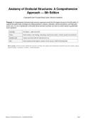
Anatomy of Orofacial Structures: A Comprehensive
Approach — 8th Edition
Expanded Exam-Focused Study Guide | Brand & Isselhard
Purpose: An independently authored study resource organized around the 36-chapter structure of the 8th edition. It
expands the public topic coverage into original questions, answers, rationales, clinical connections, and high-yield
exam cues. It is not a reproduction of the paid Stuvia test bank and does not claim to contain leaked or guaranteed
exam questions.
Coverage 36 chapters + rapid-review drills
Focus Dental anatomy, oral histology, embryology, head & neck anatomy, infection spread, local anesthesia
Question style Original exam-style Q&A with rationale/exam cue
Use Dental assisting, dental hygiene, anatomy review, quizzes, midterm/final preparation
How to study: Cover the answer, identify the structure or concept, then explain why the distractors would be wrong. For anatomy, always
connect location → structure → function → clinical relevance.
Anatomy of Orofacial Structures 8e — Expanded Study Guide Page 1
, Chapter Map
1. Oral Cavity — 10 core questions
2. The Tooth: Functions and Terms — 10 core questions
3. Fundamental and Preventive Curvatures — 10 core questions
4. Dentition — 10 core questions
5. Development, Form, and Eruption — 10 core questions
6. Occlusion — 10 core questions
7. Dental Anomalies — 10 core questions
8. Supporting Structures — 10 core questions
9. Clinical Considerations — 10 core questions
10. Tooth Identification — 10 core questions
11. Root Morphology — 10 core questions
12. Incisors — 10 core questions
13. Canines — 10 core questions
14. Premolars — 10 core questions
15. Molars — 10 core questions
16. Deciduous Dentition — 10 core questions
17. Basic Tissues — 10 core questions
18. Development of Orofacial Complex — 10 core questions
19. Dental Lamina and Enamel Organ — 10 core questions
20. Enamel, Dentin, and Pulp — 10 core questions
21. Root Formation and Attachment Apparatus — 10 core questions
22. Eruption and Shedding of Teeth — 10 core questions
23. Oral Mucous Membrane — 10 core questions
24. The Tongue — 10 core questions
25. Histology of the Salivary Glands — 10 core questions
26. Osteology of the Skull — 10 core questions
27. Nose, Nasal Cavity, and Paranasal Sinuses — 10 core questions
28. Muscles of Mastication, Hyoid Muscles, SCM and Trapezius — 10 core questions
29. Temporomandibular Joint — 10 core questions
30. Muscles of Facial Expression — 10 core questions
31. Soft Palate and Pharynx — 10 core questions
32. Arterial Supply and Venous Drainage — 10 core questions
33. Salivary Glands — 10 core questions
34. Nervous System — 10 core questions
35. Lymphatics and Spread of Dental Infection — 10 core questions
36. Anatomical Considerations in Local Anesthesia Administration — 10 core questions
Anatomy of Orofacial Structures 8e — Expanded Study Guide Page 2
, High-Yield Strategy
• Learn tooth identification by combining crown form, root morphology, arch, and distinctive landmarks.
• For head-and-neck nerves, separate sensory vs motor function and identify the division before the terminal branch.
• For local anesthesia, memorize the target nerve plus the bony/soft-tissue landmarks and nearby structures at risk.
• For infection spread, map the tooth apex relative to muscle attachments and fascial spaces, then consider airway risk.
• For histology and embryology, memorize developmental sequences rather than isolated terms.
Anatomy of Orofacial Structures 8e — Expanded Study Guide Page 3
, 1. Oral Cavity
Start with spatial orientation: vestibule, oral cavity proper, palate, tongue, and floor of mouth.
1. What is the oral vestibule?
Answer: The space between the lips or cheeks and the teeth; in an edentulous patient it lies between the lips or cheeks and the
alveolar ridges.
Exam cue: Distinguish the vestibule from the oral cavity proper.
2. What is the oral cavity proper?
Answer: The internal oral space bounded by the teeth or alveolar ridges and extending posteriorly toward the oropharyngeal
region.
Exam cue: On anatomy questions, identify the boundary being described.
3. What forms the roof of the anterior oral cavity?
Answer: The hard palate, which separates the oral cavity from the nasal cavity.
Exam cue: Hard palate = rigid anterior roof.
4. What is the soft palate?
Answer: A movable muscular fold posterior to the hard palate that contributes to swallowing and separates the nasopharynx from
the oropharynx during swallowing.
Exam cue: Soft palate questions often test swallowing and muscle function.
5. What is the floor of the mouth primarily associated with?
Answer: The tongue and muscles such as the mylohyoid, with the sublingual region containing important ducts, glands, vessels,
and nerves.
Exam cue: Know the relationship of the sublingual area to the floor of mouth.
6. What is a diastema?
Answer: A space or lack of contact between two teeth in the same dental arch.
Exam cue: A maxillary midline diastema can be associated with a prominent labial frenum.
7. What is the labial frenum?
Answer: A fold of mucosa connecting the inner lip to the alveolar mucosa or gingiva in the midline.
Exam cue: Do not confuse labial and lingual frenula.
8. What is the lingual frenum?
Answer: A mucosal fold connecting the underside of the tongue to the floor of the mouth.
Exam cue: Its anatomy can affect tongue mobility.
9. What is the gingiva?
Answer: The specialized oral mucosa surrounding and protecting the cervical portions of erupted teeth and covering alveolar bone.
Exam cue: Attached gingiva is firm; movable alveolar mucosa is different.
10. Why is oral anatomy clinically important?
Answer: Oral structures are closely related to muscles, nerves, vessels, glands, airways, and fascial spaces, so small anatomic
changes can affect function and spread of infection.
Exam cue: Think anatomy → function → clinical consequence.
2. The Tooth: Functions and Terms
Master surface terminology before attempting tooth-identification questions.
Anatomy of Orofacial Structures 8e — Expanded Study Guide Page 4
Approach — 8th Edition
Expanded Exam-Focused Study Guide | Brand & Isselhard
Purpose: An independently authored study resource organized around the 36-chapter structure of the 8th edition. It
expands the public topic coverage into original questions, answers, rationales, clinical connections, and high-yield
exam cues. It is not a reproduction of the paid Stuvia test bank and does not claim to contain leaked or guaranteed
exam questions.
Coverage 36 chapters + rapid-review drills
Focus Dental anatomy, oral histology, embryology, head & neck anatomy, infection spread, local anesthesia
Question style Original exam-style Q&A with rationale/exam cue
Use Dental assisting, dental hygiene, anatomy review, quizzes, midterm/final preparation
How to study: Cover the answer, identify the structure or concept, then explain why the distractors would be wrong. For anatomy, always
connect location → structure → function → clinical relevance.
Anatomy of Orofacial Structures 8e — Expanded Study Guide Page 1
, Chapter Map
1. Oral Cavity — 10 core questions
2. The Tooth: Functions and Terms — 10 core questions
3. Fundamental and Preventive Curvatures — 10 core questions
4. Dentition — 10 core questions
5. Development, Form, and Eruption — 10 core questions
6. Occlusion — 10 core questions
7. Dental Anomalies — 10 core questions
8. Supporting Structures — 10 core questions
9. Clinical Considerations — 10 core questions
10. Tooth Identification — 10 core questions
11. Root Morphology — 10 core questions
12. Incisors — 10 core questions
13. Canines — 10 core questions
14. Premolars — 10 core questions
15. Molars — 10 core questions
16. Deciduous Dentition — 10 core questions
17. Basic Tissues — 10 core questions
18. Development of Orofacial Complex — 10 core questions
19. Dental Lamina and Enamel Organ — 10 core questions
20. Enamel, Dentin, and Pulp — 10 core questions
21. Root Formation and Attachment Apparatus — 10 core questions
22. Eruption and Shedding of Teeth — 10 core questions
23. Oral Mucous Membrane — 10 core questions
24. The Tongue — 10 core questions
25. Histology of the Salivary Glands — 10 core questions
26. Osteology of the Skull — 10 core questions
27. Nose, Nasal Cavity, and Paranasal Sinuses — 10 core questions
28. Muscles of Mastication, Hyoid Muscles, SCM and Trapezius — 10 core questions
29. Temporomandibular Joint — 10 core questions
30. Muscles of Facial Expression — 10 core questions
31. Soft Palate and Pharynx — 10 core questions
32. Arterial Supply and Venous Drainage — 10 core questions
33. Salivary Glands — 10 core questions
34. Nervous System — 10 core questions
35. Lymphatics and Spread of Dental Infection — 10 core questions
36. Anatomical Considerations in Local Anesthesia Administration — 10 core questions
Anatomy of Orofacial Structures 8e — Expanded Study Guide Page 2
, High-Yield Strategy
• Learn tooth identification by combining crown form, root morphology, arch, and distinctive landmarks.
• For head-and-neck nerves, separate sensory vs motor function and identify the division before the terminal branch.
• For local anesthesia, memorize the target nerve plus the bony/soft-tissue landmarks and nearby structures at risk.
• For infection spread, map the tooth apex relative to muscle attachments and fascial spaces, then consider airway risk.
• For histology and embryology, memorize developmental sequences rather than isolated terms.
Anatomy of Orofacial Structures 8e — Expanded Study Guide Page 3
, 1. Oral Cavity
Start with spatial orientation: vestibule, oral cavity proper, palate, tongue, and floor of mouth.
1. What is the oral vestibule?
Answer: The space between the lips or cheeks and the teeth; in an edentulous patient it lies between the lips or cheeks and the
alveolar ridges.
Exam cue: Distinguish the vestibule from the oral cavity proper.
2. What is the oral cavity proper?
Answer: The internal oral space bounded by the teeth or alveolar ridges and extending posteriorly toward the oropharyngeal
region.
Exam cue: On anatomy questions, identify the boundary being described.
3. What forms the roof of the anterior oral cavity?
Answer: The hard palate, which separates the oral cavity from the nasal cavity.
Exam cue: Hard palate = rigid anterior roof.
4. What is the soft palate?
Answer: A movable muscular fold posterior to the hard palate that contributes to swallowing and separates the nasopharynx from
the oropharynx during swallowing.
Exam cue: Soft palate questions often test swallowing and muscle function.
5. What is the floor of the mouth primarily associated with?
Answer: The tongue and muscles such as the mylohyoid, with the sublingual region containing important ducts, glands, vessels,
and nerves.
Exam cue: Know the relationship of the sublingual area to the floor of mouth.
6. What is a diastema?
Answer: A space or lack of contact between two teeth in the same dental arch.
Exam cue: A maxillary midline diastema can be associated with a prominent labial frenum.
7. What is the labial frenum?
Answer: A fold of mucosa connecting the inner lip to the alveolar mucosa or gingiva in the midline.
Exam cue: Do not confuse labial and lingual frenula.
8. What is the lingual frenum?
Answer: A mucosal fold connecting the underside of the tongue to the floor of the mouth.
Exam cue: Its anatomy can affect tongue mobility.
9. What is the gingiva?
Answer: The specialized oral mucosa surrounding and protecting the cervical portions of erupted teeth and covering alveolar bone.
Exam cue: Attached gingiva is firm; movable alveolar mucosa is different.
10. Why is oral anatomy clinically important?
Answer: Oral structures are closely related to muscles, nerves, vessels, glands, airways, and fascial spaces, so small anatomic
changes can affect function and spread of infection.
Exam cue: Think anatomy → function → clinical consequence.
2. The Tooth: Functions and Terms
Master surface terminology before attempting tooth-identification questions.
Anatomy of Orofacial Structures 8e — Expanded Study Guide Page 4