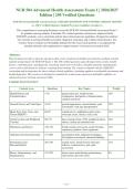
NUR 504 Advanced Health Assessment Exam 1 | 2026/2027
Edition | 250 Verified Questions
NUR 504 Advanced Health Assessment Exam 1 2026-2027 QUESTIONS AND ANSWERS ALREADY GRADED
A+. 100% Verified Solutions | Updated Per Latest Guidelines | Graded A+
This comprehensive exam prep document covers the NUR 504 Advanced Health Assessment Exam 1
for graduate nursing students. It includes 250 verified questions and answers aligned with the
2026/2027 academic year's curriculum and the latest clinical practice guidelines. Designed to reinforce
key concepts in advanced health assessment, diagnostic reasoning, and evidence-based practice, this
resource ensures students are thoroughly prepared for the exam. Each question is accompanied by
detailed rationales and explanations to support mastery of advanced assessment skills.
Abstract:
This document provides a rigorous and up-to-date review of advanced health assessment for graduate nursing
students preparing for the NUR 504 Exam 1. The 250 verified questions span all major body systems, health
history, communication, and diagnostic reasoning. Each answer includes a detailed rationale explaining the
correct choice and distractor analysis, ensuring deep learning. The content is aligned with the 2026/2027
curriculum and incorporates the latest evidence-based guidelines, including updates on telehealth assessments and
health disparities. This resource is designed to simulate the actual exam experience and build confidence in
advanced practice nursing assessment skills.
Content Area Overview:
Content Area Questions Key Topics Weight
Health History and 1-40 patient interview, health history 16%
Communication components, therapeutic communication,
cultural sensitivity
General Survey and Vital Signs 41-65 general appearance, measurements, pain 10%
assessment, vital sign interpretation
Head, Eyes, Ears, Nose, and 66-95 cranial nerves, ophthalmoscopic exam, 12%
Throat (HEENT) otoscopic exam, sinuses, oral cavity
Cardiovascular and Peripheral 96-135 heart sounds, bruits, JVD, peripheral pulses, 16%
Vascular edema, cardiac auscultation sites
Pulmonary and Thorax 136-170 breath sounds, percussion, tactile fremitus, 14%
respiratory patterns, chest expansion
Abdomen and Gastrointestinal 171-200 auscultation order, palpation techniques, 12%
liver/spleen assessment, abnormal findings
Neurological and 201-230 mental status, motor/sensory function, 12%
Musculoskeletal reflexes, cranial nerves, gait, joint
assessment
Breast, Genitourinary, and 231-250 breast exam, male/female genitourinary, 8%
Integumentary skin lesions, pressure ulcers
Page 1
,Q1. During cardiac auscultation, a low-pitched sound is heard after S2 with the patient in left lateral
decubitus. Which valvular abnormality is most likely?
A. Mitral stenosis
B. Aortic regurgitation
C. Tricuspid regurgitation
D. Pulmonary stenosis
Correct Answer: A. Mitral stenosis
Rationale: An opening snap (OS) following S2 is characteristic of mitral stenosis, resulting from abrupt
halting of the mitral valve opening due to fused commissures. Aortic regurgitation produces a diastolic
murmur, not an opening snap. Tricuspid regurgitation is systolic. Pulmonary stenosis produces a systolic
ejection murmur.
Why Wrong:
B - Aortic regurgitation produces a high-pitched decrescendo diastolic murmur, not an opening snap.
C - Tricuspid regurgitation is a holosystolic murmur heard at the left lower sternal border.
D - Pulmonary stenosis produces a systolic ejection murmur, not an opening snap.
Reference: Bickley, L.S. (2026). Bates' Guide to Physical Examination and History Taking, 14th Ed., Ch.
9
Q2. A patient's lung auscultation reveals bronchial breath sounds over the right lower lobe. Which
condition is most consistent with this finding?
A. Pleural effusion
B. Pneumothorax
C. Lobar consolidation
D. Pulmonary fibrosis
Correct Answer: C. Lobar consolidation
Rationale: Bronchial breath sounds heard over the periphery indicate consolidation (e.g., pneumonia)
where sound transmission is enhanced through solid lung tissue. Pleural effusion and pneumothorax
cause diminished or absent breath sounds. Pulmonary fibrosis typically produces fine crackles and may
have bronchial sounds only if honeycombing is severe.
Why Wrong:
A - Pleural effusion causes dullness to percussion and decreased breath sounds, not bronchial breath
sounds.
B - Pneumothorax produces hyperresonance and absent breath sounds.
D - Pulmonary fibrosis typically presents with inspiratory crackles and may have bronchial sounds
only in advanced disease with traction bronchiectasis.
Reference: Bickley, L.S. (2026). Bates' Guide to Physical Examination and History Taking, 14th Ed., Ch.
10
Page 2
,Q3. During abdominal examination, palpation of the left lower quadrant elicits pain in the right
lower quadrant. Which finding does this suggest?
A. Rovsing's sign
B. Psoas sign
C. Obturator sign
D. Cullen's sign
Correct Answer: A. Rovsing's sign
Rationale: Rovsing's sign is referred pain to the right lower quadrant when pressure is applied to the left
lower quadrant, indicative of peritoneal irritation from appendicitis. Psoas sign involves pain on right hip
extension. Obturator sign involves pain on internal rotation of the right thigh. Cullen's sign is
periumbilical ecchymosis from retroperitoneal bleeding.
Why Wrong:
B - Psoas sign is elicited by extending the right thigh with the patient in left lateral decubitus.
C - Obturator sign is elicited by internally rotating the right thigh with the hip flexed.
D - Cullen's sign is periumbilical ecchymosis, not abdominal tenderness.
Reference: Bickley, L.S. (2026). Bates' Guide to Physical Examination and History Taking, 14th Ed., Ch.
11
Q4. A patient stands with feet together and eyes closed, but sways significantly and nearly falls. The
Romberg test is positive. Which system is most likely impaired?
A. Proprioception (dorsal columns)
B. Cerebellar function
C. Labyrinthine (vestibular) function
D. Motor cortex
Correct Answer: A. Proprioception (dorsal columns)
Rationale: A positive Romberg test indicates loss of proprioceptive input from the dorsal columns; the
patient uses visual cues to compensate and sways with eyes closed. Cerebellar ataxia causes swaying even
with eyes open (Romberg is negative). Vestibular dysfunction also causes imbalance with eyes open.
Motor cortex lesions cause weakness, not sensory ataxia.
Why Wrong:
B - Cerebellar ataxia causes unsteadiness with eyes open, so Romberg test is not positive.
C - Vestibular dysfunction causes vertigo and nystagmus, not isolated swaying with eyes closed.
D - Motor cortex lesions cause contralateral weakness or paralysis, not imbalance.
Reference: Fuller, G. (2026). Neurological Examination Made Easy, 7th Ed., Ch. 6
Page 3
, Q5. A patient with shoulder pain cannot fully abduct the arm and a drop arm test is positive. Which
condition is most likely?
A. Rotator cuff tear
B. Frozen shoulder (adhesive capsulitis)
C. Subacromial bursitis
D. Glenohumeral dislocation
Correct Answer: A. Rotator cuff tear
Rationale: The drop arm test specifically assesses for full-thickness rotator cuff tear; the patient cannot
actively hold the arm abducted. Frozen shoulder presents with restricted passive and active range of
motion. Subacromial bursitis shows a painful arc but no true weakness. Dislocation causes obvious
deformity and inability to move.
Why Wrong:
B - Frozen shoulder restricts both active and passive abduction, but the drop arm test is not positive.
C - Subacromial bursitis causes pain mid-range, but often maintains strength.
D - Glenohumeral dislocation is usually traumatic with loss of shoulder contour.
Reference: Bickley, L.S. (2026). Bates' Guide to Physical Examination and History Taking, 14th Ed., Ch.
16
Q6. A patient presents with non-blanchable red-purple spots on the lower extremities. Some are
pinpoint (<2 mm), others are larger (3-5 mm). Which term correctly describes the larger lesions?
A. Petechiae
B. Purpura
C. Ecchymoses
D. Telangiectasias
Correct Answer: B. Purpura
Rationale: Purpura are non-blanchable hemorrhagic lesions 3-10 mm in diameter. Petechiae are <2 mm.
Ecchymoses are >1 cm. Telangiectasias are blanching vascular dilatations. The description of red-purple,
non-blanchable spots 3-5 mm fits purpura.
Why Wrong:
A - Petechiae are pinpoint (<2 mm), which are also present, but the larger lesions are purpura.
C - Ecchymoses are larger (>1 cm) and often darker, not 3-5 mm.
D - Telangiectasias are blanching, not non-blanchable.
Reference: Bickley, L.S. (2026). Bates' Guide to Physical Examination and History Taking, 14th Ed., Ch.
5
Page 4
Edition | 250 Verified Questions
NUR 504 Advanced Health Assessment Exam 1 2026-2027 QUESTIONS AND ANSWERS ALREADY GRADED
A+. 100% Verified Solutions | Updated Per Latest Guidelines | Graded A+
This comprehensive exam prep document covers the NUR 504 Advanced Health Assessment Exam 1
for graduate nursing students. It includes 250 verified questions and answers aligned with the
2026/2027 academic year's curriculum and the latest clinical practice guidelines. Designed to reinforce
key concepts in advanced health assessment, diagnostic reasoning, and evidence-based practice, this
resource ensures students are thoroughly prepared for the exam. Each question is accompanied by
detailed rationales and explanations to support mastery of advanced assessment skills.
Abstract:
This document provides a rigorous and up-to-date review of advanced health assessment for graduate nursing
students preparing for the NUR 504 Exam 1. The 250 verified questions span all major body systems, health
history, communication, and diagnostic reasoning. Each answer includes a detailed rationale explaining the
correct choice and distractor analysis, ensuring deep learning. The content is aligned with the 2026/2027
curriculum and incorporates the latest evidence-based guidelines, including updates on telehealth assessments and
health disparities. This resource is designed to simulate the actual exam experience and build confidence in
advanced practice nursing assessment skills.
Content Area Overview:
Content Area Questions Key Topics Weight
Health History and 1-40 patient interview, health history 16%
Communication components, therapeutic communication,
cultural sensitivity
General Survey and Vital Signs 41-65 general appearance, measurements, pain 10%
assessment, vital sign interpretation
Head, Eyes, Ears, Nose, and 66-95 cranial nerves, ophthalmoscopic exam, 12%
Throat (HEENT) otoscopic exam, sinuses, oral cavity
Cardiovascular and Peripheral 96-135 heart sounds, bruits, JVD, peripheral pulses, 16%
Vascular edema, cardiac auscultation sites
Pulmonary and Thorax 136-170 breath sounds, percussion, tactile fremitus, 14%
respiratory patterns, chest expansion
Abdomen and Gastrointestinal 171-200 auscultation order, palpation techniques, 12%
liver/spleen assessment, abnormal findings
Neurological and 201-230 mental status, motor/sensory function, 12%
Musculoskeletal reflexes, cranial nerves, gait, joint
assessment
Breast, Genitourinary, and 231-250 breast exam, male/female genitourinary, 8%
Integumentary skin lesions, pressure ulcers
Page 1
,Q1. During cardiac auscultation, a low-pitched sound is heard after S2 with the patient in left lateral
decubitus. Which valvular abnormality is most likely?
A. Mitral stenosis
B. Aortic regurgitation
C. Tricuspid regurgitation
D. Pulmonary stenosis
Correct Answer: A. Mitral stenosis
Rationale: An opening snap (OS) following S2 is characteristic of mitral stenosis, resulting from abrupt
halting of the mitral valve opening due to fused commissures. Aortic regurgitation produces a diastolic
murmur, not an opening snap. Tricuspid regurgitation is systolic. Pulmonary stenosis produces a systolic
ejection murmur.
Why Wrong:
B - Aortic regurgitation produces a high-pitched decrescendo diastolic murmur, not an opening snap.
C - Tricuspid regurgitation is a holosystolic murmur heard at the left lower sternal border.
D - Pulmonary stenosis produces a systolic ejection murmur, not an opening snap.
Reference: Bickley, L.S. (2026). Bates' Guide to Physical Examination and History Taking, 14th Ed., Ch.
9
Q2. A patient's lung auscultation reveals bronchial breath sounds over the right lower lobe. Which
condition is most consistent with this finding?
A. Pleural effusion
B. Pneumothorax
C. Lobar consolidation
D. Pulmonary fibrosis
Correct Answer: C. Lobar consolidation
Rationale: Bronchial breath sounds heard over the periphery indicate consolidation (e.g., pneumonia)
where sound transmission is enhanced through solid lung tissue. Pleural effusion and pneumothorax
cause diminished or absent breath sounds. Pulmonary fibrosis typically produces fine crackles and may
have bronchial sounds only if honeycombing is severe.
Why Wrong:
A - Pleural effusion causes dullness to percussion and decreased breath sounds, not bronchial breath
sounds.
B - Pneumothorax produces hyperresonance and absent breath sounds.
D - Pulmonary fibrosis typically presents with inspiratory crackles and may have bronchial sounds
only in advanced disease with traction bronchiectasis.
Reference: Bickley, L.S. (2026). Bates' Guide to Physical Examination and History Taking, 14th Ed., Ch.
10
Page 2
,Q3. During abdominal examination, palpation of the left lower quadrant elicits pain in the right
lower quadrant. Which finding does this suggest?
A. Rovsing's sign
B. Psoas sign
C. Obturator sign
D. Cullen's sign
Correct Answer: A. Rovsing's sign
Rationale: Rovsing's sign is referred pain to the right lower quadrant when pressure is applied to the left
lower quadrant, indicative of peritoneal irritation from appendicitis. Psoas sign involves pain on right hip
extension. Obturator sign involves pain on internal rotation of the right thigh. Cullen's sign is
periumbilical ecchymosis from retroperitoneal bleeding.
Why Wrong:
B - Psoas sign is elicited by extending the right thigh with the patient in left lateral decubitus.
C - Obturator sign is elicited by internally rotating the right thigh with the hip flexed.
D - Cullen's sign is periumbilical ecchymosis, not abdominal tenderness.
Reference: Bickley, L.S. (2026). Bates' Guide to Physical Examination and History Taking, 14th Ed., Ch.
11
Q4. A patient stands with feet together and eyes closed, but sways significantly and nearly falls. The
Romberg test is positive. Which system is most likely impaired?
A. Proprioception (dorsal columns)
B. Cerebellar function
C. Labyrinthine (vestibular) function
D. Motor cortex
Correct Answer: A. Proprioception (dorsal columns)
Rationale: A positive Romberg test indicates loss of proprioceptive input from the dorsal columns; the
patient uses visual cues to compensate and sways with eyes closed. Cerebellar ataxia causes swaying even
with eyes open (Romberg is negative). Vestibular dysfunction also causes imbalance with eyes open.
Motor cortex lesions cause weakness, not sensory ataxia.
Why Wrong:
B - Cerebellar ataxia causes unsteadiness with eyes open, so Romberg test is not positive.
C - Vestibular dysfunction causes vertigo and nystagmus, not isolated swaying with eyes closed.
D - Motor cortex lesions cause contralateral weakness or paralysis, not imbalance.
Reference: Fuller, G. (2026). Neurological Examination Made Easy, 7th Ed., Ch. 6
Page 3
, Q5. A patient with shoulder pain cannot fully abduct the arm and a drop arm test is positive. Which
condition is most likely?
A. Rotator cuff tear
B. Frozen shoulder (adhesive capsulitis)
C. Subacromial bursitis
D. Glenohumeral dislocation
Correct Answer: A. Rotator cuff tear
Rationale: The drop arm test specifically assesses for full-thickness rotator cuff tear; the patient cannot
actively hold the arm abducted. Frozen shoulder presents with restricted passive and active range of
motion. Subacromial bursitis shows a painful arc but no true weakness. Dislocation causes obvious
deformity and inability to move.
Why Wrong:
B - Frozen shoulder restricts both active and passive abduction, but the drop arm test is not positive.
C - Subacromial bursitis causes pain mid-range, but often maintains strength.
D - Glenohumeral dislocation is usually traumatic with loss of shoulder contour.
Reference: Bickley, L.S. (2026). Bates' Guide to Physical Examination and History Taking, 14th Ed., Ch.
16
Q6. A patient presents with non-blanchable red-purple spots on the lower extremities. Some are
pinpoint (<2 mm), others are larger (3-5 mm). Which term correctly describes the larger lesions?
A. Petechiae
B. Purpura
C. Ecchymoses
D. Telangiectasias
Correct Answer: B. Purpura
Rationale: Purpura are non-blanchable hemorrhagic lesions 3-10 mm in diameter. Petechiae are <2 mm.
Ecchymoses are >1 cm. Telangiectasias are blanching vascular dilatations. The description of red-purple,
non-blanchable spots 3-5 mm fits purpura.
Why Wrong:
A - Petechiae are pinpoint (<2 mm), which are also present, but the larger lesions are purpura.
C - Ecchymoses are larger (>1 cm) and often darker, not 3-5 mm.
D - Telangiectasias are blanching, not non-blanchable.
Reference: Bickley, L.S. (2026). Bates' Guide to Physical Examination and History Taking, 14th Ed., Ch.
5
Page 4